|
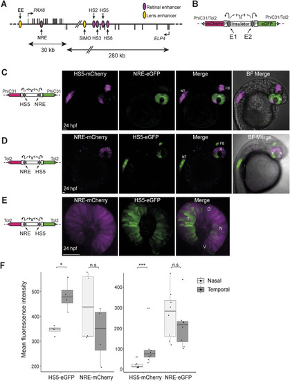
Activity of HS5 and NRE in a dual enhancer–reporter system during zebrafish embryonic development. (A) Map of human PAX6 regulatory locus showing the position of eye enhancers including HS5 and NRE (retinal, purple; lens, yellow). (B) Dual enhancer–reporter injection construct. mCherry and eGFP are transcribed from a minimal gata2 promoter (P) activated by enhancer E1 or E2. An insulator based on the chicken HS4 sequence separates the enhancers. Targeted PhiC31 integration or random Tol2 integration is used to insert the dual-reporter construct into the zebrafish genome (Bhatia et al, 2021). (C) Live imaging of a 24-hpf NRE-eGFP/HS5-mCherry F1 embryo (10x objective). NRE (eGFP) is active throughout the retina (R). HS5 (mCherry) is active in the forebrain (FB), neural tube (NT), and in the retina where activity is highest in the temporal (T) half of the retina, compared with the nasal (N) side. (D) Live imaging of a 24-hpf NRE-mCherry/HS5-eGFP F1 embryo showing the activity of HS5 (eGFP) in FB, NT, and predominantly the temporal retina, towards the ventral (V) side as opposed to dorsal (D). NRE (mCherry) is active throughout the retina (10x objective); (E, D) as in (D), but at higher resolution (40x water immersion objective). Scale bars 50 μm. (F) Quantification of mean fluorescence intensity for mCherry and eGFP in the nasal versus temporal retina at 24 hpf in NRE-mCherry/HS5-eGFP (left) and NRE-eGFP/HS5-mCherry (right) F1 embryos. The activity of HS5 (eGFP or mCherry) is significantly higher in the temporal retina. n F1 embryos imaged ≥4. Wilcoxon test results: ns, not significant; *, P < 0.05; ***, P < 0.001. Scale bars 50 μm.
|