
Fig. 7
- ID
- ZDB-FIG-230830-6
- Publication
- Spead et al., 2023 - Teneurin trans-axonal signaling prunes topographically missorted axons
- Other Figures
- All Figure Page
- Back to All Figure Page
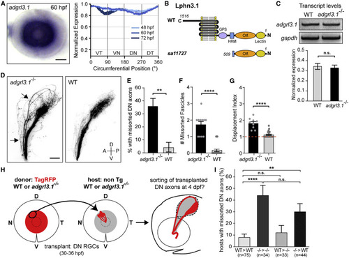
Lphn3.1 signals cell autonomously for pruning missorted DN axons (A) adgrl3.1 ISH staining is detected throughout the RGC layer in the retina throughout development. Scale bar: 50 μm. (B) Schematic of Lphn3.1 protein structure in WT and adgrl3.1sa11727 mutants. Olf, olfactomedin domain; HRM, hormone receptor motif; GPS, GPCR proteolysis site. (C) Analysis of adgrl3.1 transcript levels by RT-PCR in WT and adgrl3.1 mutants at 4 dpf. Unpaired two-tailed t test, p = 0.71. Bars: standard errors. (D) DN axons missort along the dorsal branch of the optic tract in adgrl3.1 mutants (arrows) but not in WT siblings at 4 dpf. Scale bar: 50 μm. (E) Percentage of larvae with missorted DN axons at 4 dpf. One-sided chi-squared test with Yates’s correction, X2 (1, N = 50) = 6.125, p = 0.0067. (F) Number of missorted DN axon fascicles in mutant larvae with sorting defects. Unpaired one-tailed t test, p ˂ 0.0001. (G) Quantification of the displacement index in mutant larvae with sorting defects. Unpaired one-tailed t test, p ˂ 0.0001. n = 9 mutants. (H) DN RGCs of an isl2b:TagRFP donor embryo (WT or adgrl3.1 mutant) were topographically transplanted between 30 and 36 hpf into the DN retina of a WT or adgrl3.1 mutant host. Pre-target sorting of transplanted donor DN axons was assessed at 4 dpf. (I) Quantifications of the percentage of transplanted hosts with missorted donor DN axons. Chi-squared test, X2 (3, N = 186) = 22.51, p ˂ 0.0001. ∗∗p = 0.0023; ∗∗∗∗p ˂ 0.0001. Error bars: standard errors. See also Figure S7. |