
Fig. 3
- ID
- ZDB-FIG-230830-5
- Publication
- Spead et al., 2023 - Teneurin trans-axonal signaling prunes topographically missorted axons
- Other Figures
- All Figure Page
- Back to All Figure Page
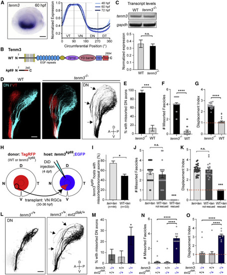
tenm3 is required for pruning topographically missorted dorsal axons (A) tenm3 ISH staining is selectively detected in the ventral RGC layer in the retina throughout development. Scale bar: 50 μm. (B) Schematic of Tenm3 protein structure in WT and tenm3kg69 mutants. Tox, toxin-like domain; YD, tyrosine-aspartate repeat; βprop, β-propeller. (C) Analysis of tenm3 transcript levels by RT-PCR in WT and tenm3 mutants at 4 dpf. Unpaired two-tailed t test p = 0.75. Error bars: standard errors. (D) Pre-target sorting of DN and VT retinal axons along the optic tract at 4 dpf. Missorted DN axons are maintained along the dorsal branch of the tract in tenm3 mutants (arrows). Scale bar: 50 μm. (E) Percentage of larvae with missorted DN axons at 4 dpf. One-sided chi-squared test with Yates’s correction, X2 (1, N = 47) = 23.30, p < 0.0001. (F) Number of missorted DN axon fascicles in phenotypic larvae. Unpaired one-tailed t test, p ˂ 0.0001. (G) Quantification of the displacement index in larvae with sorting defects. Unpaired one-tailed t test, p ˂ 0.0001, n = 20 mutants. (H) VN RGCs of an isl2b:TagRFP donor embryo (WT or tenm3 mutant) were topographically transplanted between 30 and 36 hpf into the VN retina of an isl2b:EGFP tenm3 mutant host. Pre-target sorting of host DN axons was assessed by injecting DiD into the host DN retina at 4 dpf. (I) Quantifications of the percentage of transplanted tenm3 mutant hosts with missorted DN axons. One-sided chi-squared test with Yates’s correction, X2 (1, N = 77) = 4.641, p = 0.0156. (J) Number of missorted DN axon fascicles in tenm3 mutant hosts with sorting defects. One-way ANOVA with Tukey’s post-hoc test, p ˂ 0.0001. (K) Quantification of the displacement index in tenm3 mutant hosts with sorting defects. One-way ANOVA with Tukey’s post-hoc test, p ˂ 0.0001. Error bars: standard errors. (L) Pre-target sorting of DN retinal axons along the optic tract at 4 dpf. DN axons missort along the dorsal branch of the tract in tenm3−/+; ext2dak/+ larvae (arrows) but not in tenm3−/+ heterozygotes. Scale bar: 50 μm. (M) Percentage of tenm3−/+, ext2dak/+, and tenm3−/+; ext2dak/+ larvae with missorted DN axons at 4 dpf. One-sided chi-squared test, X2 (1, N = 74) = 3.274, p = 0.035. (N) Number of missorted DN axon fascicles in phenotypic larvae. One-way ANOVA with Tukey’s post-hoc test, p ˂ 0.0001. (O) Quantification of the displacement index. One-way ANOVA with Tukey’s post-hoc test, p ˂ 0.0001. n = 25 tenm3−/+, 17 ext2dak/+, and 32 tenm3−/+; ext2dak/+. Error bars: standard errors. See also Figure S6. |