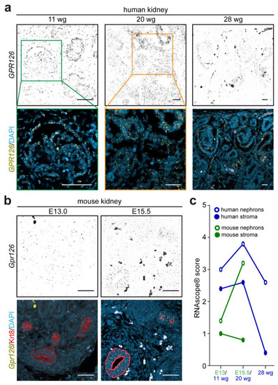
Gpr126 expression during metanephric development. (a) RNAscope® probes targeting the human GPR126 mRNA were used on human FFPE embryonic metanephric kidney sections at 11, 20, and 28 weeks of gestation (wg). Over time, the GPR126 expression pattern (yellow dotty signal) concentrates in the developing nephrons, compared to stromal cells. DAPI was utilized in (a,b) to visualize nuclei. (b) RNAscope® probes targeting the mouse Gpr126 detected low, but ubiquitously distributed, signal (yellow dotty signal) in the mouse FFPE-developing metanephros sections at E13.0 compared to samples obtained at E15.5, where signal was found more localized in cytokeratin-8-positive ureteric bud cells (red). Scale bars: 100 μm. (c) Representation of the RNAscope® signal score means obtained from epithelial (circumferences) and stromal (circles) cell types of human (blue) and mouse (green) developing metanephroi during the physiological nephrogenesis burst window (20 wg in human, n = 5; E15.5 in mouse, n = 3), earlier (11 wg in human, n = 4; E13.0 in mouse, n = 3), and later (28 wg in human, n = 3) stages.
|
