FIGURE

Figure 4.

ID
ZDB-FIG-230606-23
Publication
Hamling et al., 2023 - The Nature and Origin of Synaptic Inputs to Vestibulospinal Neurons in the Larval Zebrafish
Other Figures
All Figure Page
Back to All Figure Page
Figure 4.

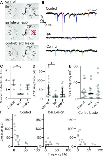
High-amplitude spontaneous excitatory inputs originate in the ipsilateral ear. A, Lesion schematic: the utricle (gray circle) was physically removed (red “x”) either ipsilateral or contralateral to the recorded vestibulospinal neuron (black circle, “VS”). B, Example current traces from neurons held at −75 mV from control (top), ipsilateral (middle), and contralateral (bottom) experiments; EPSCs are in color. C, Number of EPSC amplitude bins per cell [median ± interquartile range (IQR) in black] is decreased after ipsilateral, but not contralateral, lesion. Asterisks indicate statistically significant differences (p < 0.05). D, EPSC bin amplitudes (median ± IQR in black) are decreased after ipsilateral, but not contralateral, lesion. E, Frequency of events in EPSC bins (median ± IQR in black) is unchanged after ipsilateral or contralateral lesion compared with control cells. F, EPSC amplitude versus frequency for each bin in control and after ipsilateral/contralateral lesions. High-amplitude bins are lost after ipsilateral lesion.

Expression Data

Expression Detail
Antibody Labeling
Phenotype Data

Phenotype Detail
Acknowledgments
This image is the copyrighted work of the attributed author or publisher, and ZFIN has permission only to display this image to its users. Additional permissions should be obtained from the applicable author or publisher of the image. Full text @ eNeuro