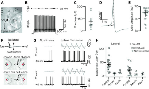
Vestibulospinal neurons encode utricle-derived body translation. A, Schematic of spinal-projecting vestibulospinal (VS) neuron targeted for electrophysiology. B, Vestibulospinal membrane potential at rest (top, 0 pA) and in response to current injection (bottom, 98 pA). C, Rheobase (mean ± SEM) across 12 nonspontaneously active cells. D, Example action potential waveform with amplitude (dotted line). E, Action potential average amplitude (median ± interquartile range) across 21 cells. F, Immobilized fish were manually translated in the fore–aft or lateral axes (top). Vestibulospinal neurons were recorded in control and after two manipulations: first, in otogelin mutants (middle) that do not develop utricles (red “x”) and second, after chemically induced hair cell (red “x”) death (bottom). G, Accelerometer (gray) and voltage trace (black) from a neuron in a control fish (top) showing action potentials in phase with translation. In contrast, activity from an otogelin mutant (bottom) is unaligned with translation. H, Modulation depth of spiking response (mean ± SEM) is disrupted in both the lateral (left) and fore–aft (right) directions after both chronic and acute disruption of the utricle. Gray circles, neurons; black outlined circles, statistically significant directional responses.
|
