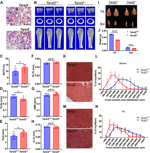
Tarsl2 deletion affects the development of bone and muscle.A, H&E staining of bone sections from 8-week-old Tarsl2+/+ and Tarsl2−/− mice. The black arrow indicates the abnormal trabeculae in the tibia of the Tarsl2−/− mice. The scale bar represents 40 mm. B, three-dimensional μ-CT images of the trabecular bone in distal femurs isolated from 8-week-old male Tarsl2+/+ and Tarsl2−/− mice (n = 6). C–H, the μ-CT results in (B) were quantitatively analyzed to determine bone volume fraction (BV/TV), trabecular thickness (Tb.Th), the number of trabeculae (TB.N), the separation of trabeculae (Tb.Sp), bone mineral density (BMD), and cortical bone thickness (Ct.Th) (n = 6). I, comparisons of the tibialis anterior muscle and soleus muscle between the 6-week-old Tarsl2+/+ and Tarsl2−/− mice. The scale bar represents 1 cm. J, the weight of the TA and soleus muscle of the Tarsl2+/+ and Tarsl2−/− mice in (I) was measured. K, H&E staining of soleus cross sections from 6-week-old Tarsl2+/+ and Tarsl2−/− mice. L, area of the soleus cross section from Tarsl2+/+ and Tarsl2−/− mice. M, H&E staining of TA cross sections from 6-week-old Tarsl2+/+ and Tarsl2−/− mice. N, area of TA cross sections of Tarsl2+/+ and Tarsl2−/− mice. ∗∗∗∗p < 0.0001, ∗∗∗p < 0.001, and ∗p < 0.05. The error bar represents the mean ± SD with the p values indicated (two-tailed Student’s t test). TA, tibialis anterior.
|
