
Fig. 8
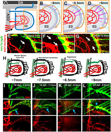
Late-stage remodeling leading to the formation of the interplexus bridges and regression of the dorsal arm of the primary arc. (A-D) Schematics illustrating the formation of links between proximal and distal vascular plexuses. DA, dorsal aorta; ED, endoskeletal disk. (A) Simplified overview diagram of the pectoral fin vasculature at ∼3.5 weeks, showing the proximal plexus (red) and arterial feed for the distal fin-ray vascular network (orange). Orange box in A shows the approximate magnified area depicted in B-D. (B-D) Stages in the formation of inter-plexus links. (E-G) Confocal images of the inter-plexus region of the pectoral fin of three separate 3.5-week-old Tg(kdrl:mcherry)y206, Tg(mrc1a:egfp)y251 double-transgenic zebrafish, showing the formation of arterial [Tg(kdrl:mcherry)y206 positive, Tg(mrc1a:egfp)y251 negative] links (white arrows) between the medial proximal plexus (left) and the arterial fin-ray vascular plexus (right). (H) Schematics of the replacement of the dorsal arm of the primary arc. (I-L) Confocal images of the dorsal arm of the pectoral fin primary arc showing the early Tg(mrc1a:egfp)y251-positive, Tg(kdrl:mcherry)y206-negative sprout (I), the elongation of the sprout toward the distal plexus (J), two lumenized connections leading to the CCV (K), and the regression of the original dorsal arm of the primary arc, leaving only the new, deeper connection (L). Cyan arrows emphasize the new venous sprout in each panel. Dashed lines indicate the common cardinal vein. Images shown in E-G and I-L are representative of data collected from 12 separate larvae. Scale bars: 50 μm. |