
Fig. 4
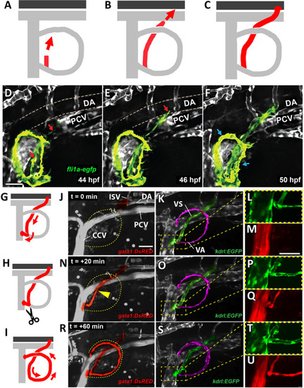
Perfusion of the primitive pectoral artery. (A-C) Schematics illustrating stages in the formation of a connecting link between the primitive pectoral artery and the dorsal aorta, including formation of a new sprout from the base of the primitive pectoral artery where it connects ventrally to the CCV (A), medial and dorsal-caudal elongation of the new vessel segment (B), and linkage to the dorsal aorta at or near the base of the second ISV (C). (D-F) Confocal still images from a time-lapse series showing formation of a connecting link between the primitive pectoral artery and the dorsal aorta in a Tg(fli1a:egfp)y1 transgenic zebrafish at 44 hpf (D), 46 hpf (E) and 50 hpf (F). Red arrows indicate the migratory direction of the arterial sprout, and blue arrows indicate perfusion of the primary arc. See Movie 2 for the complete time-lapse series. (G-I) Schematics illustrating initial shunting of flow from the dorsal aorta through the base of the pectoral fin directly to the CCV (G), followed by disconnection of this shunt (H) and re-routing of flow through the primitive pectoral artery primary arc (I). (J-U) Confocal images from a time-lapse series of the pectoral fin and adjacent structures in a 2-day-old Tg(gata1:dsred)sd2Tg, Tg(kdrl:egfp)s843 double-transgenic zebrafish, showing Tg(gata1:dsred)sd2Tg-positive blood cells (J,M,N,Q,R,U) and Tg(kdrl:egfp)s843-positive blood vessels (K,L,O,P,S,T). L, P and T show higher-magnification images of the boxed areas in K, O and S, respectively; likewise, M, Q and U are higher-magnification images of the corresponding areas in J, N and R, respectively. (J-M) t=0 min. The vascular link to the dorsal aorta has assembled (highlighted green in K) but has not yet lumenized, and no blood flow is present. (N-Q) t=+20 min. The connection to the dorsal aorta (highlighted green in O) has been completed and blood is now flowing through this connecting vessel (highlighted red in N and Q), but the blood is being shunted directly to the CCV without perfusing the primitive pectoral artery arc (highlighted magenta in K,O,S). (R-U) t=+60 min. The shunt to the CCV has disconnected and all blood flow has been rerouted into the primitive pectoral artery arc (highlighted red in R and U). Blood is only emptying into the CCV through the dorsal connection. See Movie 4 for the complete time-lapse series. See Movies 5 and 6 for higher-magnification images and 3D reconstructions of the ventral junction to the CCV and its shunting and then disconnection. Images shown are representative of data collected from 11 separate embryos. Yellow arrowhead in N indicates red blood cells flowing through the arterial sprout, and dashed yellow line encircles primary arc in J,N,R. DA, dorsal aorta; PCV, posterior cardinal vein; VA, ventral arm of the primary arc; VS, ventral arterial sprout. Scale bars: 30 μm (D-F); 40 μm (J-U). |