
Fig. 1
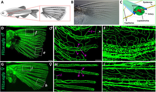
Vascular anatomy of the adult zebrafish pectoral fin. (A) Drawing of an adult zebrafish with a magnified view of the pectoral fin and its lepidotrichia (fin-ray bones). (B) Transmitted light image of a dissected pectoral fin. (C) Schematic showing a cross-section of a zebrafish fin ray. (D-I) Tiled confocal images of pectoral fins dissected from adult male (D-F) or adult female (G-I) Tg(fli1a:egfp)y1 transgenic zebrafish. E and F show higher-magnification views of the boxed regions in D. H and I show higher-magnification views of the boxed regions in G. a, fin-ray artery; i, fin-ray interlinking vessels; v, paired fin-ray veins. The asterisk in E denotes an artifact of image stitching. Images shown are representative of data collected from three separate male and three separate female adult fins, with consistent observation of the overall patterning of major vessels. Scale bars: 500 μm (B,D,G); 150 µm (E,H); 100 µm (F,I). |