
FIGURE
Fig. 8
- ID
- ZDB-FIG-220912-99
- Publication
- Atienzar-Aroca et al., 2022 - Transgenic Overexpression of Myocilin Leads to Variable Ocular Anterior Segment and Retinal Alterations Associated with Extracellular Matrix Abnormalities in Adult Zebrafish
- Other Figures
- All Figure Page
- Back to All Figure Page
Fig. 8
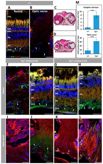
Figure 8. Retinal and optic nerve gliosis in old (two years) myocilin transgenic zebrafish. A rabbit anti-GFAP primary antibody was used to detected Müller glial cells. (A) Wild-type retina and (B) optic nerve. (C,D) Representative transgenic head tissue sections indicating the regions analysed by immunohistochemistry. (E–H) Transgenic retina. (I–L) Transgenic optic nerve. (M) Quantification of GFAP-positive cells in the ganglion cell layer and optic nerve. Four microscopic fields per eye were analyzed (n = four eyes). ***: p < 0.001, Student’s t-test. Arrowheads: GFAP labelling of Müller cells. Scale bars: 50 μm. Autofl.: tissue autofluorescence used for image contrast and anatomical reference; GCL: ganglion cell layer; IPL: inner plexiform layer; INL: inner nuclear layer; OA: ocular alterations; OPL: outer plexiform layer; ONL; outer nuclear layer; PHL: photoreceptor layer; R: retina. Tg/+: transgenic; +/+: wild type. The images are representative of the results observed in two zebrafish of each type.
|
Expression Data
| Antibody: | |
|---|---|
| Fish: | |
| Anatomical Terms: | |
| Stage: | Adult |
Expression Detail
Antibody Labeling
Phenotype Data
| Fish: | |
|---|---|
| Observed In: | |
| Stage: | Adult |
Phenotype Detail
Acknowledgments
This image is the copyrighted work of the attributed author or publisher, and
ZFIN has permission only to display this image to its users.
Additional permissions should be obtained from the applicable author or publisher of the image.
Full text @ Int. J. Mol. Sci.