
FIGURE
Fig. 6
- ID
- ZDB-FIG-220912-97
- Publication
- Atienzar-Aroca et al., 2022 - Transgenic Overexpression of Myocilin Leads to Variable Ocular Anterior Segment and Retinal Alterations Associated with Extracellular Matrix Abnormalities in Adult Zebrafish
- Other Figures
- All Figure Page
- Back to All Figure Page
Fig. 6
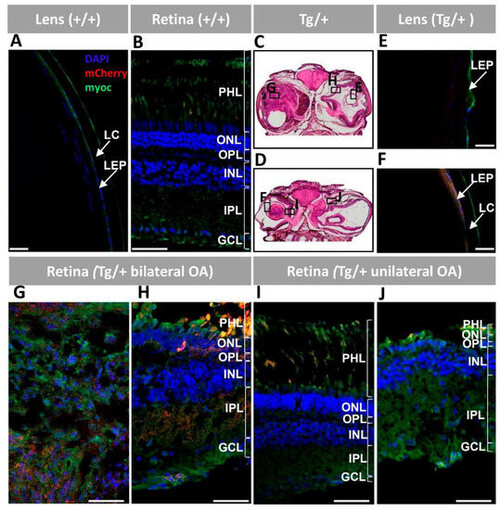
Figure 6. Fluorescent immunohistochemistry expression analysis of myocilin and mCherry in the lens and retina of old (two-year-old) myoc transgenic zebrafish. A chicken anti-myocilin primary antibody was used to detected myocilin (green signal). Control wild type lens (A) and retina (B). Representative head tissue sections in panels (C,D) indicate the regions analyzed by immunohistochemistry in transgenic zebrafish with bilateral (C) or unilateral (D) ocular alterations. Lens (E,F) and retina (G–J) tissue sections. Scale bars: 50 μm. GCL: ganglion cell layer; IPL: inner plexiform layer; INL: inner nuclear layer; LC: lens capsule; LEP: lens epithelium; OA: ocular alterations; OPL: outer plexiform layer; ONL; outer nuclear layer; PHL: photoreceptor layer; +/+: wild type; Tg/+: transgenic.
|
Expression Data
Expression Detail
Antibody Labeling
Phenotype Data
Phenotype Detail
Acknowledgments
This image is the copyrighted work of the attributed author or publisher, and
ZFIN has permission only to display this image to its users.
Additional permissions should be obtained from the applicable author or publisher of the image.
Full text @ Int. J. Mol. Sci.