
FIGURE
Fig. 13
- ID
- ZDB-FIG-220912-104
- Publication
- Atienzar-Aroca et al., 2022 - Transgenic Overexpression of Myocilin Leads to Variable Ocular Anterior Segment and Retinal Alterations Associated with Extracellular Matrix Abnormalities in Adult Zebrafish
- Other Figures
- All Figure Page
- Back to All Figure Page
Fig. 13
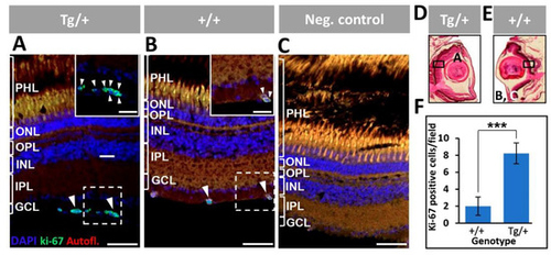
Figure 13. Proliferation of retinal ganglion cells in adult (13-month-old) myocilin transgenic zebrafish. (A,B) An anti-Ki-67 primary antibody was used to detect proliferating cells. Digital magnification of the positive cells indicted with white dotted boxes are shown in the inserts. (C) The negative control consisted of tissue sections incubated only with the secondary antibody. (D,E) Black rectangles in two representative eye sections indicate the location of microscopic fields shown in the different panels. (F) Quantification of Ki-67-positive cells. Four microscopic fields per eye were analyzed (n = four eyes). ***: p < 0.001, Student’s t-test. Scale bars: 50 μm. Arrowheads: Ki-67-positive cells. Scale bars: 50 μm. GCL: ganglion cell layer; IPL: inner plexiform layer; INL: inner nuclear layer; OPL: outer plexiform layer; ONL: outer nuclear layer; PHL: photoreceptor layer. The images are representative of the results observed in two fishes of each genotype.
|
Expression Data
Expression Detail
Antibody Labeling
Phenotype Data
| Fish: | |
|---|---|
| Observed In: | |
| Stage: | Adult |
Phenotype Detail
Acknowledgments
This image is the copyrighted work of the attributed author or publisher, and
ZFIN has permission only to display this image to its users.
Additional permissions should be obtained from the applicable author or publisher of the image.
Full text @ Int. J. Mol. Sci.