
Fig. 6
- ID
- ZDB-FIG-220801-227
- Publication
- Gurung et al., 2022 - Single-cell transcriptomic analysis of vascular endothelial cells in zebrafish embryos
- Other Figures
- All Figure Page
- Back to All Figure Page
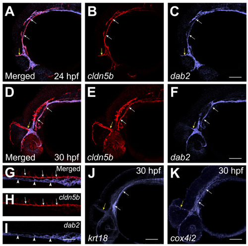
Vascular endothelial (VE)-other cluster #6 corresponds to cells in cranial vasculature. (A–F) Two color fluorescent ISH analysis for the expression of cldn5b and dab2 at the 24 hpf and 30 hpf stages. Note that cldn5b and dab2 are co-expressed in the PHBC (white arrows) and MCeV (yellow arrows) at the 24 hpf and 30 hpf stages. (G–I) Two color fluorescent ISH analysis for the expression of arterial cldn5b and venous dab2 in the trunk region of 24 hpf embryos. Note that cldn5b and dab2 have distinct non-overlapping expression in the DA (arrows) and PCV (arrowheads) respectively. (J,K) Fluorescent ISH analysis for the expression of krt18 and cox4i2 in the head region of 30 hpf embryos. Both markers are expressed in the PHBC (white arrows) and MCeV (yellow arrows). PHBC primordial hindbrain channels, MCeV mid cerebral vein, DA dorsal aorta, PCV posterior cardinal vein. Scale bars: 100 μm. |
| Genes: | |
|---|---|
| Fish: | |
| Anatomical Terms: | |
| Stage Range: | Prim-5 to Prim-15 |