
Fig. 3
- ID
- ZDB-FIG-220623-39
- Publication
- Georgantzoglou et al., 2022 - A two-step search and run response to gradients shapes leukocyte navigation in vivo
- Other Figures
- All Figure Page
- Back to All Figure Page
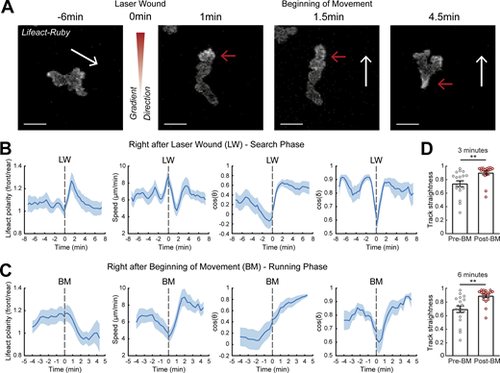
A two-stage response to newly encountered gradients in vivo. (A) Time-lapse sequence of two-photon confocal image projections showing a neutrophil (white) in a Tg(mpx:Lifeact-Ruby) zebrafish larva, migrating pre- and post-LW. Cell movement started at 1.5-min after laser wounding for this example cell. White arrows indicate the speed vector. Red arrows indicate the side of cell with higher Lifeact abundance. Scale bar = 10 μm. (B and C) Neutrophil Lifeact polarity, speed, cosine of θ, and cosine of δ, in relation to time, respectively. Time sequence was synchronized based on time of the LW (B) or the time that each individual neutrophil began to move post-LW (C). (B)n = 21 cells, from 9 larvae. (C)n = 18 cells, from 9 larvae. (D) Neutrophil track straightness for motion completed within 3 (top) or 6 (bottom) min before and after beginning of neutrophil movement (BM). n = 18 cells, from 9 larvae. Mean and SEM are shown. Wilcoxon matched-pairs signed rank test, **, P = 0.0040 (3 min), P = 0.0019 (6 min). |