
Fig. 1
- ID
- ZDB-FIG-220623-37
- Publication
- Georgantzoglou et al., 2022 - A two-step search and run response to gradients shapes leukocyte navigation in vivo
- Other Figures
- All Figure Page
- Back to All Figure Page
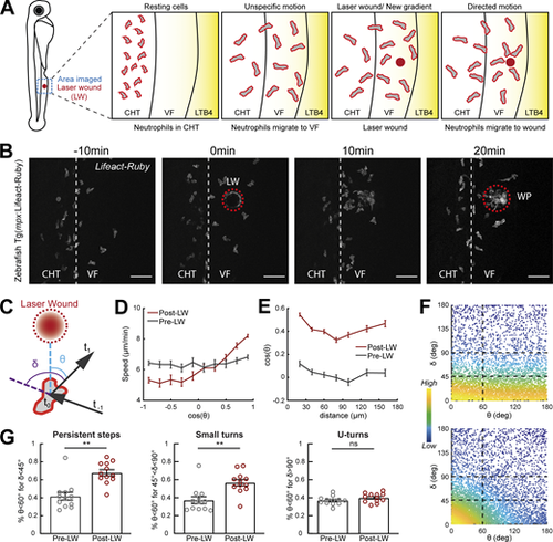
Gradients in vivo cause a directional bias on speed and persistence and small reorientations towards the source. Analysis of neutrophil migration responses to gradients in zebrafish. (A) Scheme showing two-photon LW chemotaxis assay in zebrafish. Resting neutrophils at the CHT are stimulated to exit the VF after LTB4 addition. Laser wounding in the VF at 30 min after LTB4 addition redirects neutrophil motion. (B) Time-lapse sequence of two-photon confocal image projections, showing fluorescent neutrophils in 3 dpf Tg(mpx:Lifeact-Ruby) transgenic larva, migrating pre- and post-LW (red dashed line). LW occurs at 0 min. Scale bar = 50 μm. (C) Scheme for angles δ and θ. δ is the angle between the speed vector of three successive timepoints (t0–t−1, t1–t0). θ is the angle between the speed vector (t1–t0) and the vector connecting the neutrophil geometrical center to the closest point of the LW perimeter. (D) Cell speed in relation to the cosine of θ for pre- and post-LW. n = 343–1,283 cell steps per bin pre-wound, n = 236–3,168 cell steps per bin post-wound. (E) Cell cosine of θ in relation to the distance from the closest point of the WP. n = 517–863 cell steps per bin for pre-wound, n = 444–1,272 cell steps per bin for post-wound. (F) Scatter plot of cell δ in relation to θ, color-coded for the density of points, for pre- (top) and post-LW (bottom). Black dashed lines indicate the groups of data points taken for analysis for G. Data shown from 12 larvae. (G) Percentage of cell persistent steps (δ < 45°), small turns (45° < δ < 90°) and U- turns (δ > 90°), respectively, with θ < 60°. Wilcoxon matched-pairs signed rank test, **, P = 0.0024 (persistent steps), P = 0.0068 (small turns). (D, E, and G) Data from 12 larvae in each case. |