
FIGURE
Fig. 5
- ID
- ZDB-FIG-220504-20
- Publication
- Meng et al., 2021 - Cardiac toxicity assessment of pendimethalin in zebrafish embryos
- Other Figures
- All Figure Page
- Back to All Figure Page
Fig. 5
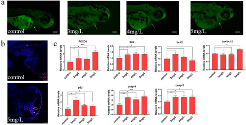
PND induced apoptosis of zebrafish cells. (a) AO staining. The green highlights represent apoptotic cells; Scale = 350 µm. (b) Representative images of TUNEL-stained embryos showing apoptotic cells (white arrow). (c) After exposure to PND, the transcription level of apoptosis related genes was changed. The values are expressed as the mean ± standard deviation of three independent experiments (n = 3). *p < 0.05, **p < 0.01, ***p < 0.001. |
Expression Data
| Genes: | |
|---|---|
| Fish: | |
| Condition: | |
| Anatomical Term: | |
| Stage: | Protruding-mouth |
Expression Detail
Antibody Labeling
Phenotype Data
| Fish: | |
|---|---|
| Condition: | |
| Observed In: | |
| Stage: | Protruding-mouth |
Phenotype Detail
Acknowledgments
This image is the copyrighted work of the attributed author or publisher, and
ZFIN has permission only to display this image to its users.
Additional permissions should be obtained from the applicable author or publisher of the image.
Full text @ Ecotoxicol. Environ. Saf.