|
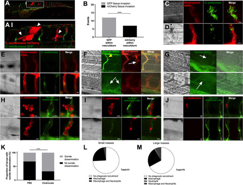
Dissemination events through vasculature damage.A-B Infection of 2 dpf AB larvae with 25 cfu of a 5:1 ratio of GFP:mCherry KN99 C. neoformans. Larvae were serieally imaged until 8 dpf, or death A-AI white arrow heads indicate examples of dissemination of C. neoformans (mCherry) into the somite surrounding an existing mCherry cryptococcal mass B Comparison of colour of C. neoformans in the vasculature (GFP or mCherry), and the corresponding colour of dissemination events at the same location (19 events in total) C, D and E Infection of KDRL mCherry blood marker transgenic line at 2 dpf with 1000 cfu GFP C. neoformans C Dissemination from an intact blood vessel, with C. neoformans in the surrounding tissue suggested to be transcytosis D Damaged blood vessels with C. neoformans in surrounding tissue E Intact blood vessels (KDRL marker) with or without C. neoformans F-G Infection of KDRL mCherry blood marker transgenic line at 2 dpf with 1000 cfu C. neoformans and then injection of 40kDa FITC Dextran (green) at 5 dpf immediately before imaging F yellow arrowhead indicates dextran within unblocked inter-segmental vessel, white arrows indicate cryptococcal masses within inter-segmental vessels which do not have dextran within. G white arrows indicate sites of dextran leakage into surrounding somite next to cryptococcal dissemination events. H-J Infection of vascular-endothelium cadherin endothelial junction (blood vessel marker) transgenic line with 1000 cfu mCherry C. neoformans H Intact blood vessel endothelial layer, with C. neoformans in the surrounding tissue I Damage in the blood vessel endothelial layer J Intact blood vessels with or without C. neoformans K Proportion of larvae developing cryptococcal somite growths by 3 dpi, infected with 500 cfu H99-GFP C. neoformans with clodronate liposome or PBS-control treatment (n = 3, groups of 92 and 145 larvae). L-M Infection of Tg(mpeg1:mCherry.CAAX)sh378 stably crossed to Tg(mpx:eGFP)i114 larvae at 2 dpf with 1000 cfu KN99 C. neoformans imaged at 3 dpi L The number of small cryptococcal masses (up to 4 cryptococcal cells) with macrophage, neutrophil, both or no phagocyte recruitment (n = 3, 26 larvae) M The number of larger cryptococcal masses (over 4 cryptococcal cells) with macrophage, neutrophil, both or no phagocyte recruitment (n = 3, 26 larvae). In this figure images of vessels show a single plane of a 50 μm stack.
|