|
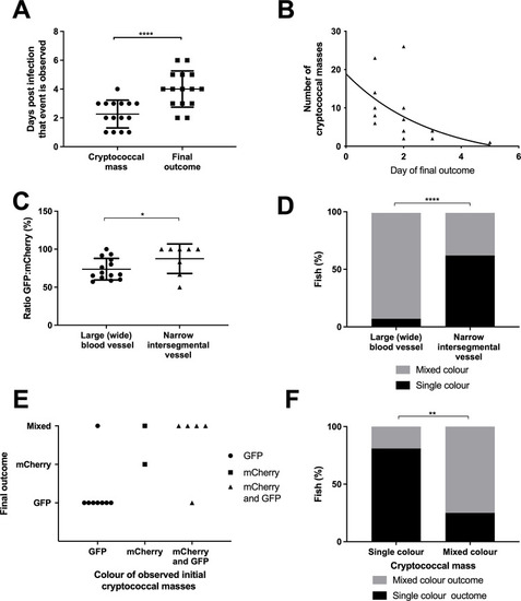
Cryptococcal mass formation leads to uncontrolled infection.Infection of 2 dpf AB larvae with 25 cfu of a 5:1 ratio of GFP:mCherry KN99 C. neoformans. Larvae were imaged until 8 dpf, or death (n = 3, in each repeat 7, 10 and 12 larvae were used) A Time cryptococcal mass first observed and time of final outcome observed (n = 3, +/- SEM, Wilcoxon matched pairs test, ****p<0.0001) B The maximun number of cryptococcal masss observed within individual larvae and how many days after observation final overwhelming infection was reached (n = 3, non-linear regression, one-phase decay) C The ratio of GFP:mCherry C.neoformans in the large caudal vein in comparison to the fifth intersegmental blood vessel, at uncontrolled infection time point (n = 3, *p<0.05, +/-SEM, paired t-test). D Single or mixed C.neoformans strains in the large caudal vein in comparison to the fifth inter-segmental blood vessel, at uncontrolled infection time point (n = 3, ****p<0.0001, Fischer’s exact test). E Comparison of the colour (either GFP, mCherry or mixed) of C. neoformans in cryptococcal masses found in inter-segmental vessels, in relation to the final outcome majority C. neoformans colour F Comparison of the colour of cryptococcal masss, either single colour or mixed, with the colour of final outcome (n = 3, **p<0.01, Fischer’s exact test).
|