|
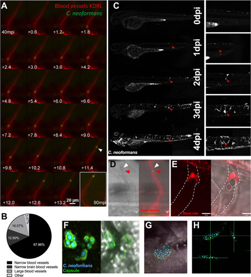
Cryptococcal mass formation by cryptococcal cell trapping in small blood vessels in the zebrafish.A Infection of KDRL mCherry blood marker transgenic line with 25 cfu GFP C. neoformans, imaged immediately after infection. A single cryptococcal cell becomes trapped in the vasculature (white arrow), at 40 minutes post infection (mpi) after moving from the bottom of the vessel toward the top (left to right, time points +0.6 seconds). Last image shows cryptococcal cell in the same location at the end of the time-lapse at 90mpi B Infection of 2 dpf AB larvae with 25 cfu of a 5:1 ratio of GFP:mCherry KN99 C. neoformans. Larvae were imaged until 8 dpf, or death (n = 3, in each repeat 7, 10 and 12 larvae were used) Proportion of cryptococcal masss observed in small intersegmental blood vessels, small brain blood vessels, large caudal vein or in other locations e.g. yolk, (n = 3). C Infection of 2 dpf AB larvae with 25 cfu of a 5:1 ratio of GFP:mCherry KN99 C. neoformans. Larvae were imaged until 8 dpf, or death (n = 3, in each repeat 7, 10 and 12 larvae were used). In this case an mCherry majority overwhelming infection was reached. Infection progression from 0 dpi (day of infection imaged 2 hpi), until 4 dpi. Red arrows follows an individual cryptococcal mass formation and ultimate dissemination. D Infection of 2 dpf AB larvae with 25 cfu of a 5:1 ratio of GFP:mCherry KN99 C. neoformans showing blood cells (white arrow) trapped behind a cryptococcal mass (red arrow) within an inter-segmental vessel. E Infection of 2 dpf Tg(gata1:dsRed) larvae with 1000 cfu GFP of KN99 C. neoformans showing blood cells (red) trapped behind a cryptococcal mass within an inter-segmental vessel (white dashed lines). F-H GFP KN99 (cyan), antibody labelled cryptococcal capsule (green). F Cryptococci within blood vessels demonstrating the enlarged capsule blocking the vessel 24 hpi G-H Cryptococcal mass encased in capsule. G Merged florescence and transmitted light z projection H Three-dimensional section of cryptococcal mass showing encasement in polysaccharide capsule.
|