
Fig. 2
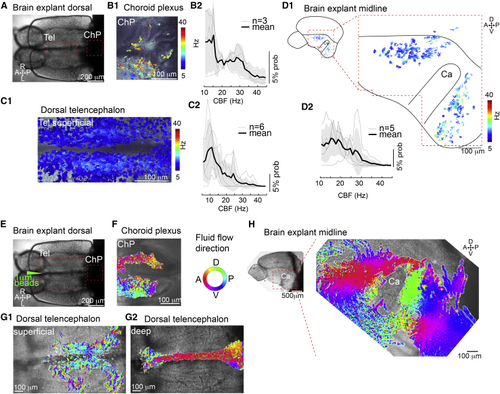
Cilia in the telencephalon, TC, and ChP are motile and generate CSF flow (A) Dorsal view of an adult brain explant. (B–D) Cilia in the forebrain ChP (B), TC (C), and telencephalic midline (D) are motile as shown by analysis of high-speed video microscopy using a pixel-based Fourier analysis. (B1–D1) Map of ciliary beating with ciliary beat frequency (CBF) color-coded in the ChP (B1), TC (C1), and hemisphere explant anterior and posterior to the Ca (D1). (B2–D2) Probability histogram showing the frequency of the pixel-based analysis and the average ± SEM for ChP (B2), TC (C2), and telencephalic hemisphere (D2). n = number of brain explants. (E) Dorsal view of an adult brain explant injected with 1 μm fluorescent beads. (F–H) Directional fluid flow in the ChP (F; n = 4), dorsal telencephalon (G; n = 5), and telencephalic midline (H; n = 5) is color-coded. Tel, telencephalon; A, anterior; P, posterior; D, dorsal; V, ventral; R, right; L, left. See also |