
Figure 7
- ID
- ZDB-FIG-211029-165
- Publication
- Ando et al., 2021 - Zebrafish Vascular Mural Cell Biology: Recent Advances, Development, and Functions
- Other Figures
- All Figure Page
- Back to All Figure Page
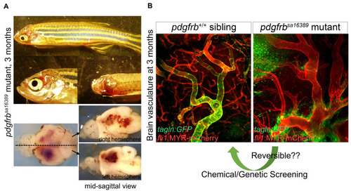
Abnormal vascular formation in pdgfrb mutant. (A) pdgfrbsa16389 mutants have no apparent vascular abnormalities, edema, or signs of hemorrhage during early developmental stages [53]. They start to show clear vascular abnormalities, however, after 1 month of age. After the juvenile stage, they have signs of bleeding (arrows) and the associated prominent phenotype in brain vessels. Images of brains dissected from the fish shown on the top are presented in the lower panels. (B) Comparison of the brain vasculature of wild-type (left) and pdgfrbsa16389 mutant (right) mice, showing the same anatomical region, confirms the capillary network reduction and dilation of arteries. Coverage with VSMC visualized by TgBAC(tagln:EGFP)ncv25Tg reporter (green) found in the wild type is absent in the pdgfrb mutant. Regardless of how severe vascular defects are, pdgfrb mutant zebrafish can reach adulthood and produce viable offspring, which is a unique feature of zebrafish and indicates the utility of the pdgfrb mutant zebrafish for chemical or genetic screening, allowing the discovery of treatments for vascular anomalies such as aneurysms. Scale bars: 50 μm (B). Fluorescent images are from [53]. |