
Figure 6
- ID
- ZDB-FIG-211029-164
- Publication
- Ando et al., 2021 - Zebrafish Vascular Mural Cell Biology: Recent Advances, Development, and Functions
- Other Figures
- All Figure Page
- Back to All Figure Page
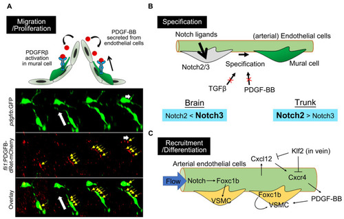
Molecular mechanisms underlying zebrafish mural cell development. (A) PDGF-BB/PDGFRβ signaling is essential for zebrafish mural cell migration and proliferation. The timelapse imaging of brain mural cells during the embryonic stage, at the bottom, shows the uptake of mCherry-fused PDGF-B into the mural cell processes (yellow arrows) extending toward the direction of migration. White arrows indicate the direction of mural cell migration. mCherry-fused PDGF-B depleted of the retention motif was originally expressed by endothelial cells using the fli1 promoter. This observation fits the proposed model in which PDGF-BB secreted from endothelial cells activates PDGFRβ expressed in mural cells to attract these cells to the vascular wall and the leading front. (B) Notch2/3 are both indispensable for mural cell specification, with a preference for Notch3 in the brain and Notch2 in the trunk. TGFβ or PDGF-BB is not essential for specification into mural cells, at least during early developmental stages, in the brain or trunk vessels of zebrafish. (C) Arterial endothelial cells induce VSMC recruitment or VSMC differentiation via Foxc1b or in a PDGF-BB-dependent manner. Cell-autonomous Foxc1b function, in VSMC developing from progenitors, has also been reported. |