
Figure 3
- ID
- ZDB-FIG-211029-161
- Publication
- Ando et al., 2021 - Zebrafish Vascular Mural Cell Biology: Recent Advances, Development, and Functions
- Other Figures
- All Figure Page
- Back to All Figure Page
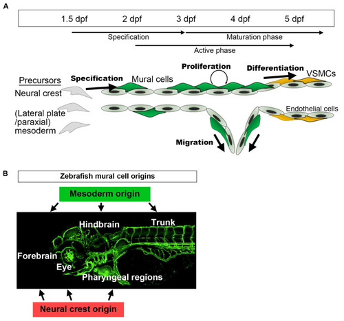
A schematic presentation of the developmental time course of zebrafish mural cells mainly in the brain and trunk. (A) Neural crest or mesoderm-derived mural cell precursors located in the vicinity of (arterial) endothelial cells are destined for the mural cell lineage during the period 36–72 hpf. Subsequently, such mural cells actively proliferate and migrate to cover arterial vessels. This proliferation and migration of mural cells appears to take place when active angiogenesis is induced (active phase). Along with the establishment of a vascular hierarchy, mural cells on larger caliber vessels start to differentiate into VSMCs after approximately 72 hpf (maturation phase). Upon formation of the vascular system, mural cell proliferation, migration, and differentiation take place. Mural cells, other than those on the dorsal/ventral aorta, resemble pericytes when they first appear, especially in the brain. However, whether they are identical to pericytes or still undergoing maturation (progenitor) to differentiate into VSMCs or pericytes remains unknown. (B) Mural cells in the hindbrain and trunk are of mesoderm origin, and those in pharyngeal regions and eyes are from the neural crest. The anterior part of the brain (forebrain) contains mural cells derived from both the mesoderm and the neural crest [12]. |