FIGURE

Figure 1

ID
ZDB-FIG-211029-159
Publication
Ando et al., 2021 - Zebrafish Vascular Mural Cell Biology: Recent Advances, Development, and Functions
Other Figures
All Figure Page
Back to All Figure Page
Figure 1

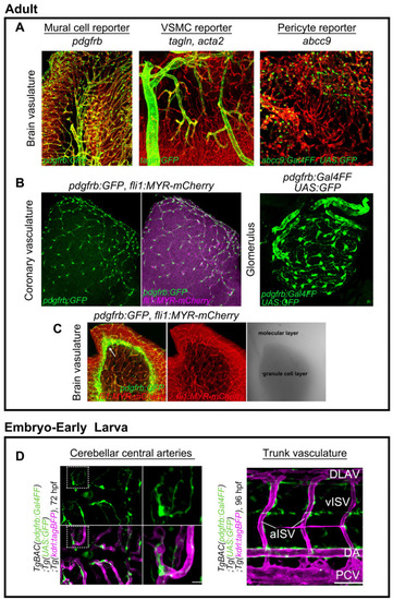
Transgenic zebrafish fluorescent reporter lines for mural cells. (A) Expressions of TgBAC(pdgfrb:GFP)ncv22Tg (left), TgBAC(tagln:EGFP)ncv25Tg (center), and TgBAC(abcc9:GAL4FF)ncv34Tg (right) reporters in zebrafish adult brain. Vessels are labeled with Tg(fli1:MYR-mCherry)ncv1Tg or Tg(kdrl:DsRed2)pd27 (red). All types of mural cells are labeled with pdgfrb. VSMCs can be labeled with tagln or acta2 reporter. Brain pericytes are selectively labeled with the abcc9 reporter [2]. See also Table 1. (B) reporter labels mural cells in coronary vessels and glomerulus mesangial cells. (C) Non-vascular-associated pdgfrb expression (green) at the interface between the molecular and granule cell layers. Images are transverse sectional views of the adult zebrafish cerebellum. (D) pdgfrb reporter (green) labels mural cells in the brain and trunk from the early stages of development. Scale bars: 50 μm, 20 μm (enlarged images).

Expression Data

Expression Detail
Antibody Labeling
Phenotype Data

Phenotype Detail
Acknowledgments
This image is the copyrighted work of the attributed author or publisher, and ZFIN has permission only to display this image to its users. Additional permissions should be obtained from the applicable author or publisher of the image. Full text @ Life (Basel)