FIGURE

FIGURE 4

ID
ZDB-FIG-210902-28
Publication
Koh et al., 2021 - Fluorescence Correlation Spectroscopy Reveals Survival Motor Neuron Oligomerization but No Active Transport in Motor Axons of a Zebrafish Model for Spinal Muscular Atrophy
Other Figures
All Figure Page
Back to All Figure Page
FIGURE 4

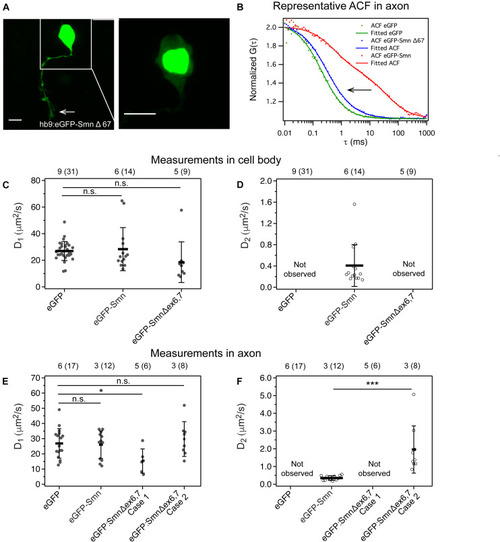
Expression and dynamics of eGFP- Smn Δex6,7 in zebrafish motor neurons. (A) Expression of eGFP- SmnΔex6,7 in motor neuron cell body and axon at 2 dpf. It is expressed strongly in the nucleus and weakly in axon and cytoplasm of cell body. (B) Representative ACF and fitted ACF of eGFP, eGFP-Smn and eGFP-SmnΔex6,7. (C) Average diffusion coefficients, D1 (μm2/s) of eGFP, eGFP-Smn, eGFP-SmnΔex6,7 in cell body. (D) Average diffusion coefficients, D2 (μm2/s) of eGFP-Smn, eGFP-SmnΔex6,7 in cell body. (E) Average diffusion coefficients, D1 (μm2/s) of eGFP, eGFP-Smn, eGFP- SmnΔex6,7 in axons. (F) Average diffusion coefficients, D2 (μm2/s) of eGFP-Smn, eGFP-SmnΔex6,7 in axons. Error bars represent the SD. (n.s.P > 0.05, P < 0.05, ∗∗∗P < 0.001). Measurements grouped under Case 1 were fitted to 3D 1-particle diffusion model, found in 6 out of 14 measurements, indicating a loss of the second component. Case 2 refers to measurements fitted to 3D 2-particle diffusion model, found in 8 out of 14 measurements. The numbers above the graph indicate the numbers of fish (number of points) the measurements were taken in.

Expression Data

Expression Detail
Antibody Labeling
Phenotype Data

Phenotype Detail
Acknowledgments
This image is the copyrighted work of the attributed author or publisher, and ZFIN has permission only to display this image to its users. Additional permissions should be obtained from the applicable author or publisher of the image. Full text @ Front Cell Dev Biol