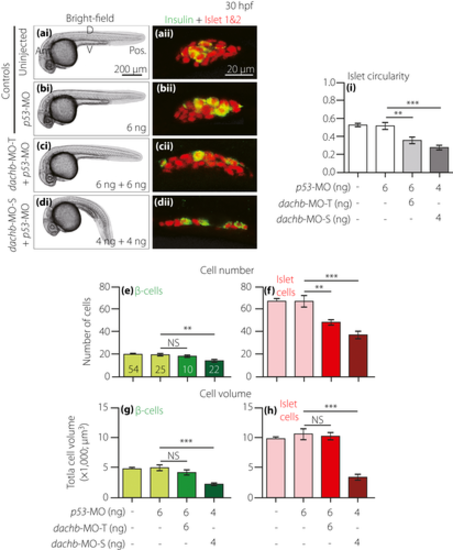
Comparison of dachb knockdown with translation-blocking morpholino (dachb-MO-T) and splice-blocking dachb-MO (dachb-MO-S) on the development of the pancreatic islet. Embryos were (a) uninjected (control) or they were injected at the one-cell stage with (b) 6 ng p53-MO (control), (c) 6 ng dachb-MO-T + 6 ng p53-MO or (d) 4 ng dachb-MO-S + 4 ng p53-MO. (ai–di) Bright-field images were acquired at 30 hpf, after which the embryos were whole-mount double-immunolabeled with antibodies to insulin (green) and islet 1 and 2 (red) to show the localization of the β-cells and islet cells in the endocrine pancreas, respectively. (aii–dii) These are all single optical sections taken through the middle of the pancreatic islet. (e,f) The number of (e) β-cells and (f) islet cells, and (g,h) the volume of (g) β-cells and (h) islet cells. (i) The circularity of the pancreatic islet. The data are presented as the mean ± standard error of n = 10 to 54 embryos, such that the number is shown in the respective bars in panel (e). The asterisks indicate significant differences at P < 0.01** or P < 0.001*** using the Mann–Whitney U-test. Ant., anterior; D, dorsal; NS, not significant; Pos. posterior; V, ventral.
|
