
Figure 2
- ID
- ZDB-FIG-210606-161
- Publication
- Rohrer et al., 2021 - Conditional Loss of the Exocyst Component Exoc5 in Retinal Pigment Epithelium (RPE) Results in RPE Dysfunction, Photoreceptor Cell Degeneration, and Decreased Visual Function
- Other Figures
- All Figure Page
- Back to All Figure Page
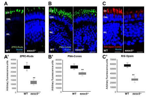
Immunohistochemical analysis of rod and cone photoreceptors in wild-type and exoc5−/− mutant zebrafish. Rod photoreceptor outer segments were identified with ZPR3 antibody (green, ZPR3, (A)), cone photoreceptors with PNA-lectin-488 (green, PNA, (B)), and medium-wavelength R/G cone opsins (red, R/G Opsin, (C)), all at 3.5 dpf. Loss of rod and cone OS immunofluorescence was noted in exoc5−/− mutant zebrafish. Scale bars = 50 μm (B) and 25 μm (A,C). OS, outer segments; ONL, outer nuclear layer; INL, inner nuclear layer. (A’–C’) Image J was used to quantify immunofluorescence for the 3 OS markers. ** p < 0.005 (WT vs. exoc5−/− mutants). |
| Antibody: | |
|---|---|
| Fish: | |
| Anatomical Term: | |
| Stage: | Protruding-mouth |
| Fish: | |
|---|---|
| Observed In: | |
| Stage: | Protruding-mouth |