
|
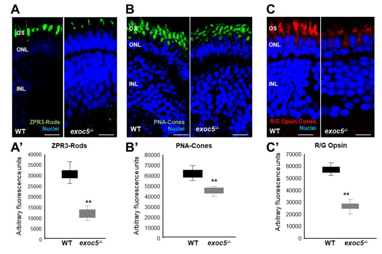
Figure 2 Immunohistochemical analysis of rod and cone photoreceptors in wild-type and exoc5−/− mutant zebrafish. Rod photoreceptor outer segments were identified with ZPR3 antibody (green, ZPR3, (A)), cone photoreceptors with PNA-lectin-488 (green, PNA, (B)), and medium-wavelength R/G cone opsins (red, R/G Opsin, (C)), all at 3.5 dpf. Loss of rod and cone OS immunofluorescence was noted in exoc5−/− mutant zebrafish. Scale bars = 50 μm (B) and 25 μm (A,C). OS, outer segments; ONL, outer nuclear layer; INL, inner nuclear layer. (A’–C’) Image J was used to quantify immunofluorescence for the 3 OS markers. ** p < 0.005 (WT vs. exoc5−/− mutants).