
- Title
-
Conditional Loss of the Exocyst Component Exoc5 in Retinal Pigment Epithelium (RPE) Results in RPE Dysfunction, Photoreceptor Cell Degeneration, and Decreased Visual Function
- Authors
- Rohrer, B., Biswal, M.R., Obert, E., Dang, Y., Su, Y., Zuo, X., Fogelgren, B., Kondkar, A.A., Lobo, G.P., Lipschutz, J.H.
- Source
- Full text @ Int. J. Mol. Sci.
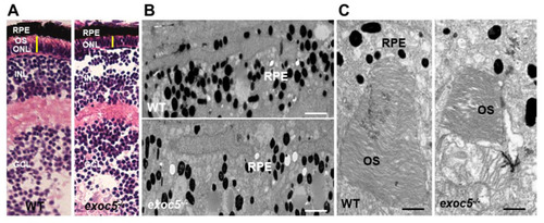
Histological and TEM analysis of RPE and retinas of wild-type and exoc5 mutant zebrafish larvae. (A) In systemic exoc5 homozygous mutants (exoc5−/−), the photoreceptor outer segments (OS) were shorter compared to wild-type (WT) siblings. (B) Ultrastructural analysis of WT and exoc5−/− mutant RPE using transmission electron microscopy indicate reduced levels of melanosomes in the mutant fish RPE. (C) WT photoreceptors showed tightly stacked disk membranes, while in exoc5−/− mutants, only remnants of outer segments (OS) could be observed. Please note the apparent formation of ectosomes at the base of the mutant photoreceptor OS. Scale bars = 2 μm (B), 800 nm (C). OS, outer segments; ONL, outer nuclear layer; RPE, retinal pigmented epithelium; INL, inner nuclear layer; GCL, ganglion cell layer. |
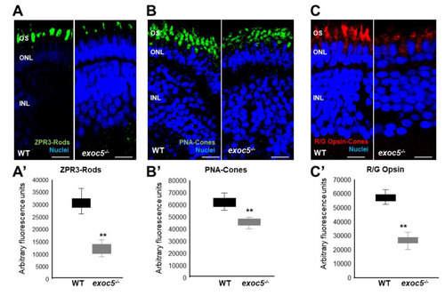
Immunohistochemical analysis of rod and cone photoreceptors in wild-type and exoc5−/− mutant zebrafish. Rod photoreceptor outer segments were identified with ZPR3 antibody (green, ZPR3, (A)), cone photoreceptors with PNA-lectin-488 (green, PNA, (B)), and medium-wavelength R/G cone opsins (red, R/G Opsin, (C)), all at 3.5 dpf. Loss of rod and cone OS immunofluorescence was noted in exoc5−/− mutant zebrafish. Scale bars = 50 μm (B) and 25 μm (A,C). OS, outer segments; ONL, outer nuclear layer; INL, inner nuclear layer. (A’–C’) Image J was used to quantify immunofluorescence for the 3 OS markers. ** p < 0.005 (WT vs. exoc5−/− mutants). EXPRESSION / LABELING:
PHENOTYPE:
|
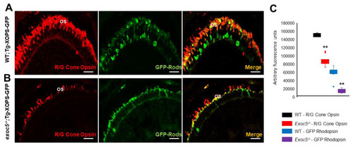
Immunohistochemical analysis of rod and cone photoreceptors in transgenic (Tg:XOPS) wild-type and exoc5 mutant zebrafish. Top panel: wild-type:Transgenic-XOPS (WT;Tg-XOPS-GFP) zebrafish retinas immunostained with R/G cone opsin (red). Bottom panel: exoc5−/− mutant:Transgenic-XOPS (exoc5−/−;Tg-XOPS-GFP) retinas immunostained with R/G cone opsin (red). The transgenic line expresses soluble endogenous GFP in rods (green). Significantly shorter rod and cone OS were observed in exoc5−/− mutant zebrafish, compared to WT zebrafish, at similar ages. (C) Image J was used to quantify fluorescence for R/G cone opsin and GFP+ rods. ** p < 0.005 (WT vs. exoc5−/− mutants). EXPRESSION / LABELING:
PHENOTYPE:
|
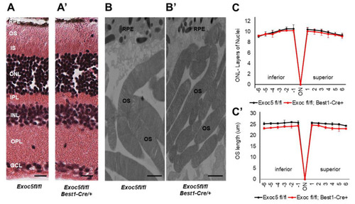
Histological and TEM analysis of retinas of Exoc5−/− mutants and WT mice at 20 weeks of age. (A,A’) H&E staining of WT and Exoc5−/− retinas at 20 weeks did not reveal histological differences. (B,B’) Transmission electron microscopy of wild-type (WT) mice photoreceptor cells show that rod OSs are shorter and thinner in Exoc5−/− retinas. The thickness of the ONL (C) and OS lengths (C’) from H&E sections through the optic nerve (ON; 0 μm distance from Optic Nerve and the starting point) was measured at 12 locations around the retina, six each in the superior and inferior hemispheres, each equally at 150 μm distances. (B,B’) Scale bars = 800 nm. OS, outer segments; RPE, retinal pigmented epithelium; IS, inner segments; ONL, outer nuclear layer; INL, inner nuclear layer; OPL, outer plexiform layer. |

Histological and TEM analysis of retinas of Exoc5−/− mutants and WT mice at 27 weeks of age. (A,A’) H&E staining of WT and Exoc5−/− retinas at 27 weeks suggests the presence of disorganized rod outer segments (OS). (B,B’) Transmission electron microscopy of WT and Exoc5−/− photoreceptor cells reveal packages of rod OS in the subretinal space. (C,C’) Thickness of the ONL (C) and OS lengths (C’) from H&E sections through the optic nerve (ON; 0 μm distance from Optic Nerve and starting point) was measured at 12 locations around the retina, six each in the superior and inferior hemispheres, each equally at 150 μm distances. Scale bars = 800 nm (B,B’). OS, outer segments; RPE, retinal pigmented epithelium; IS, inner segments; ONL, outer nuclear layer; INL, inner nuclear layer; OPL, outer plexiform layer. |
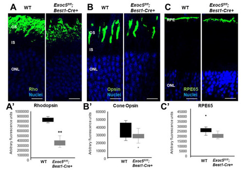
Immunohistochemical analysis of rod and cone photoreceptors in wild-type and conditional |
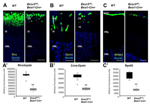
Immunohistochemical analysis of rod and cone photoreceptors in wild-type and conditional Exoc5 knockout mice at 27 weeks of age. Levels and localization of rhodopsin (green, Rho, (A)), Red/Green cone opsins (green, R/G Opsin, (B)), and RPE65 (green, RPE65, (C)), were assessed using immunohistochemistry in retinal sections from mice collected at 27 weeks of age to identify alterations in rod and cone outer segments and RPE. Rod and cone outer segments are dysmorphic, and RPE staining for RPE65 was severely disrupted. Image J was used to quantify rhodopsin (A’) and cone opsin (B’) fluorescence in photoreceptors, and RPE65 (C’) in RPE of WT and Exoc5fl/fl;Best1-Cre+ mice. Scale bars = 50 μm (A–C). OS, outer segments; IS, inner segments; ONL, outer nuclear layer, INL, inner nuclear layer; RPE, retinal pigmented epithelium. ** p < 0.05 (WT vs. Exoc5fl/fl;Best1-Cre+ mutants). |
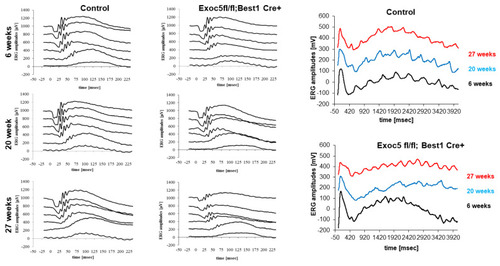
Measurement of visual function in Exoc5fl/fl;Best1-Cre+ mice by full-field electroretinography (ERG). Dark-adapted, scotopic ERGs were recorded in response to increasing light intensities in cohorts of 6, 20, and 27-week old control (Exoc5fl/fl) and Exoc5fl/fl;Best1-Cre+ mice. RPE-specific Exoc5 knockout mice in which both copies of Exoc5 were eliminated showed progressively significantly lower dark-adapted a- and b-wave amplitudes compared to controls (posthoc ANOVA: a-waves p < 0.03; b-waves p < 0.01), in particular at higher light intensities (20, 10, 6, 0 dB). C-waves showed an age-dependent decline in amplitude in the Exoc5 fl/fl;Best1-Cre+ mice (p = 0.002). Data are expressed as mean ± SEM (Exoc5 fl/fl;Best1-Cre-: n = 10; and Exoc5 fl/fl;Best1-Cre+ mice: n = 10). |