|
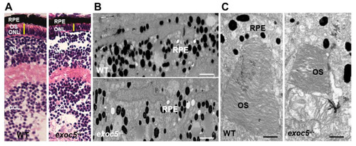
Histological and TEM analysis of RPE and retinas of wild-type and exoc5 mutant zebrafish larvae. (A) In systemic exoc5 homozygous mutants (exoc5−/−), the photoreceptor outer segments (OS) were shorter compared to wild-type (WT) siblings. (B) Ultrastructural analysis of WT and exoc5−/− mutant RPE using transmission electron microscopy indicate reduced levels of melanosomes in the mutant fish RPE. (C) WT photoreceptors showed tightly stacked disk membranes, while in exoc5−/− mutants, only remnants of outer segments (OS) could be observed. Please note the apparent formation of ectosomes at the base of the mutant photoreceptor OS. Scale bars = 2 μm (B), 800 nm (C). OS, outer segments; ONL, outer nuclear layer; RPE, retinal pigmented epithelium; INL, inner nuclear layer; GCL, ganglion cell layer.
|