
Figure 8
- ID
- ZDB-FIG-210606-167
- Publication
- Rohrer et al., 2021 - Conditional Loss of the Exocyst Component Exoc5 in Retinal Pigment Epithelium (RPE) Results in RPE Dysfunction, Photoreceptor Cell Degeneration, and Decreased Visual Function
- Other Figures
- All Figure Page
- Back to All Figure Page
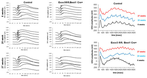
Measurement of visual function in Exoc5fl/fl;Best1-Cre+ mice by full-field electroretinography (ERG). Dark-adapted, scotopic ERGs were recorded in response to increasing light intensities in cohorts of 6, 20, and 27-week old control (Exoc5fl/fl) and Exoc5fl/fl;Best1-Cre+ mice. RPE-specific Exoc5 knockout mice in which both copies of Exoc5 were eliminated showed progressively significantly lower dark-adapted a- and b-wave amplitudes compared to controls (posthoc ANOVA: a-waves p < 0.03; b-waves p < 0.01), in particular at higher light intensities (20, 10, 6, 0 dB). C-waves showed an age-dependent decline in amplitude in the Exoc5 fl/fl;Best1-Cre+ mice (p = 0.002). Data are expressed as mean ± SEM (Exoc5 fl/fl;Best1-Cre-: n = 10; and Exoc5 fl/fl;Best1-Cre+ mice: n = 10). |