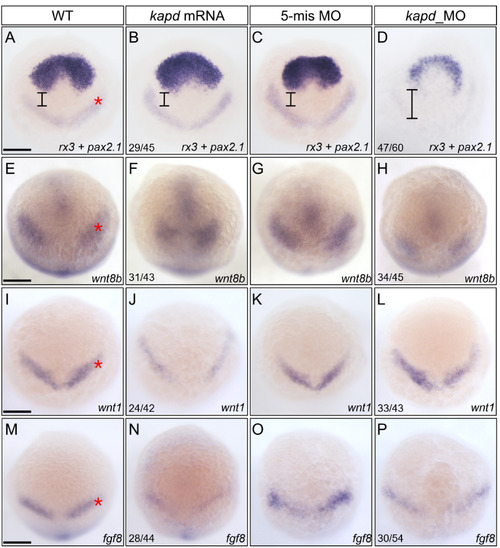
Spatiotemporal expression patterns of markers for presumptive midbrain and DA progenitors at 10 hpf . (A-D) WISH analysis using rx3 and pax2.1 as a molecular marker for the forebrain and midbrain domains indicates that loss of kapd function disrupted the presumptive midbrain development. (A) WT embryo. Microinjection of kapd mRNA (30 pg per embryo) into wild-type embryos thickened the anterior forebrain (B). kapd 5-mismatch control embryos had the similar expression patterns in the presumptive midbrain (C) to that of WT (A). In contrast, knock-down of kapd (0.2 ng morpholino per embryo) repressed MHB (asterisk) and telencephalic precursors in kapd morphants at 10 hpf (D). (E-H) WISH analysis with wnt8b as a molecular marker for the midbrain in anterior brain patterning. wnt8b transcripts were present in the midbrain of WT (E) and control embryos injected with 5-mismatch (G). Overexpression of kapd (kapd mRNA 30 pg per embryo) shortened the MHB area (F) while knock-down of kapd (0.2 ng of kapd morpholino per embryo) remarkably reduced the level of wnt8b transcripts in the MHB (asterisk) and anterior midbrain of the morphants (H). (I-P) WISH analysis with wnt1 and fgf8 as molecular markers for DA progenitors at 10 hpf. WT embryos (I and M), embryos injected with kapd mRNA (J and N). kapd mRNA injected embryos showed narrower and reduced expression patterns for wnt1 (J) and fgf8 (N). The level of wnt1 transcripts was not significantly changed in the midbrain neural keel region of kapd morphants (L). However, transcripts of fgf8 were reduced in the MHB neural keel in comparison to those of control embryos at 10 hpf (M-P). (A-P) Scale bars = 250 μm.
|
