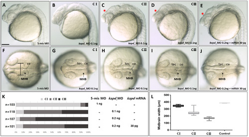
Repression of kapd expression decreases the size of the mesencephalon, including the midbrain ventricle, at 24 hpf. Microinjection of kapd morpholino (0.1 ng) into an embryo at the one-cell stage to knock down DEG-13. (A) 5-mismatch MO and (B-D) kapd morphants were categorized as class I, II, or III (C I, C II, or C III). (E) Rescue of a kapd morphant (0.2 ng) with kapd mRNA (30 pg). (F-J) Dorsal view of the midbrain, midbrain–hindbrain boundary, and hindbrain of kapd morphants at 24 hpf. (F) 5-mismatch MO. (G-I) At 24 hpf, embryos were injected with kapd MO. (J) kapd mRNA (30 pg) was injected into a kapd morphant at the one-cell stage (0.2 ng). (K) The proportion of moderately deformed embryos (class II) was higher after rescue with kapd mRNA. (L) Measurement of midbrain widths revealed a significant reduction of the neural tube in kapd morphants. Statistical significance was calculated using one-way ANOVA, and multiple comparisons between groups were analyzed by Dunnett’s test. (A-J) Scale bar = 250 µm. MHB, midbrain–hindbrain boundary; ce, cerebellum; tec, tectum.
|
