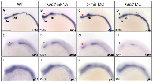
Knockdown of kapd expression alters the expression patterns of the molecular markers shha and shhb. (A-D) WISH analysis using shha as a molecular marker for the thalamus (asterisks) and zona limitans intrathalamica (ZLI) revealed that loss of kapd function disrupts diencephalic differentiation. (A) WT embryo. Microinjection of kapd mRNA (50 pg per embryo) into WT embryos increased expression in the hypothalamus (hy) (B). Expression patterns of kapd in the hypothalamus of 5-mismatch MO control embryos (C) were similar to those of WT embryos (A). By contrast, in kapd morphants at 16.5 hpf, knockdown of kapd (0.2 ng MO per embryo) decreased expression in the hypothalamus (D). (E-H) shhb transcripts were present in the ventral FP (asterisks) of WT embryos at 16.5 hpf (E). Overexpression of kapd did not cause significant changes in the ventral FP of the forebrain (F), whereas knockdown of kapd (0.2 ng of kapd morpholino per embryo) markedly decreased the level of shhb transcripts in the ventral diencephalon of the morphants (H). Embryos were examined for expression of the ventral neural marker, gli1, at 16.5 hpf. Transcript levels of gli1 were not affected by overexpression or knockdown of kapd (I-L). (A-L) Scale bar = 250 µm.
|
