
Figure 5
- ID
- ZDB-FIG-210422-23
- Publication
- Rho et al., 2021 - Protocol for analysis of integrin-mediated cell adhesion of lateral plate mesoderm cells isolated from zebrafish embryos
- Other Figures
- All Figure Page
- Back to All Figure Page
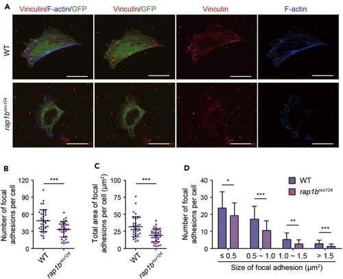
Rap1b regulates integrin-mediated adhesion of LPM cells to fibronectin-coated dishes (A) Cells dissociated from 17 hpf wild type (upper) and (B) Number of vinculin-marked focal adhesions per cell, as observed in (A). (C) Total area of vinculin-marked focal adhesions per cell, as observed in (A). (D) Number of vinculin-marked focal adhesions, as observed in (A). Data are shown as means ± s.d. ∗p < 0.05; ∗∗p < 0.01; ∗∗∗p < 0.001. (B)-(D) reuse parts of a figure from Rho et al. ( |