
FIGURE
Figure 3
- ID
- ZDB-FIG-210422-21
- Publication
- Rho et al., 2021 - Protocol for analysis of integrin-mediated cell adhesion of lateral plate mesoderm cells isolated from zebrafish embryos
- Other Figures
- All Figure Page
- Back to All Figure Page
Figure 3
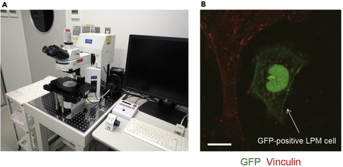
LPM cells adhering to the fibronectin-coated dish (A) FluoView FV1000 confocal upright microscope system. (B) The cells derived from 17 hpf embryos were left to adhere to the fibronectin-coated dish for 13 h and then stained with anti-GFP (green) and anti-vinculin (red) antibodies. Arrow indicates GFP-positive LPM cell. Scale bar, 10 μm. |
Expression Data
Expression Detail
Antibody Labeling
Phenotype Data
Phenotype Detail
Acknowledgments
This image is the copyrighted work of the attributed author or publisher, and
ZFIN has permission only to display this image to its users.
Additional permissions should be obtained from the applicable author or publisher of the image.
Full text @ STAR Protoc