
Figure 2
- ID
- ZDB-FIG-210422-20
- Publication
- Rho et al., 2021 - Protocol for analysis of integrin-mediated cell adhesion of lateral plate mesoderm cells isolated from zebrafish embryos
- Other Figures
- All Figure Page
- Back to All Figure Page
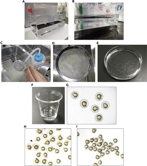
Preparation of the dechorionated zebrafish embryos at 17 hpf (A) Set up zebrafish to breed in the separate breeding tank (step 1). (B) Initiate breeding by removing the divider separating the male and female fish (step 2). (C) Rinse the embryos on the strainer with 0.03% sea salt solution (step 5). (D) Cleaned embryos on the strainer. (E) The embryos transferred into a 10-cm petri dish containing E3 medium (step 6). (F and G) The 17 hpf embryos transferred into a beaker containing E3 medium (step 8b). (H) The embryos treated with the Pronase solution (step 8d). Note that the chorions start to break open (arrow). (I) Dechorionated embryos in a 10-cm petri dish containing E3 medium (step 8g). |