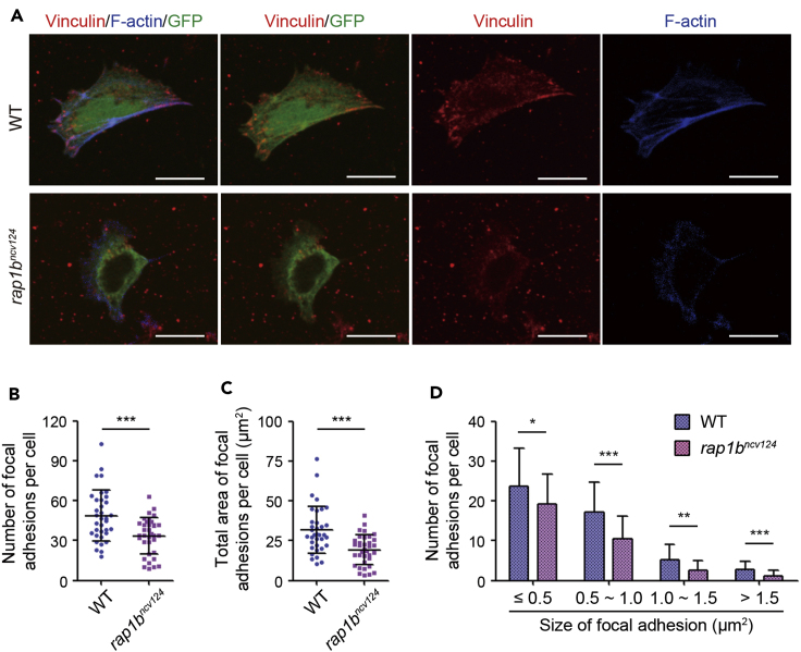
IMAGE

Figure 5

ID
ZDB-IMAGE-210422-23
Source
Figures for Rho et al., 2021
Image
Figure Caption

Figure 5

Rap1b regulates integrin-mediated adhesion of LPM cells to fibronectin-coated dishes

(A) Cells dissociated from 17 hpf wild type (upper) and rap1bncv124 (lower) Tg(fli1a:GFP)y1 embryos. Merged images of GFP (green)/vinculin (red)/F-actin (blue), those of GFP (green)/vinculin (red), and vinculin (red) and F-actin (blue) images are shown, as indicated. Scale bars, 10 μm.

(B) Number of vinculin-marked focal adhesions per cell, as observed in (A).

(C) Total area of vinculin-marked focal adhesions per cell, as observed in (A).

(D) Number of vinculin-marked focal adhesions, as observed in (A). Data are shown as means ± s.d. ∗p < 0.05; ∗∗p < 0.01; ∗∗∗p < 0.001. (B)-(D) reuse parts of a figure from Rho et al. (Rho et al., 2019).

Acknowledgments
This image is the copyrighted work of the attributed author or publisher, and ZFIN has permission only to display this image to its users. Additional permissions should be obtained from the applicable author or publisher of the image. Full text @ STAR Protoc