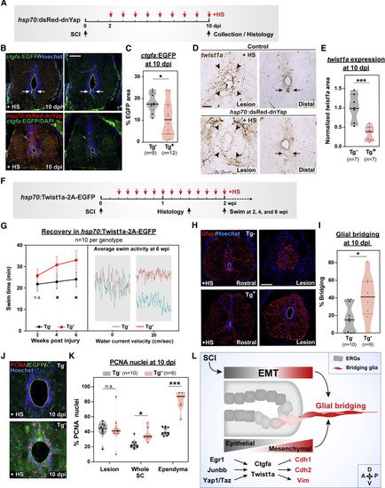
Yap signaling directs twist1a-driven EMT and ctgfa-dependent glial bridging (A–C) ctgfa reporter expression in ctgfa:EGFP;hsp70:dsRed-dnYap (Tg+) SCs. ctgfa:EGFP (Tg−) siblings were used as controls. All animals were subjected to SCI and daily heat shocks (+HS) (A). EGFP expression was assessed at 10 dpi (B). Dotted lines delineate central canal edges. For quantification, the area of EGFP fluorescence was calculated for 2 sections per animal (C). (D and E) twist1a expression in hsp70:dsRed-dnYap and control wild-type siblings. All animals were subjected to daily heat shocks (+HS). RNAscope was performed at 10 dpi (D). Arrowheads point to the lesion core; arrows point to ventral ependymal progenitors in distal SC sections. For quantification, normalized twist1a area was calculated for 2 sections per animal (E). (F and G) Swim assays determined motor function recovery of hsp70:Twist1a-2A-EGFP (Tg+, red) and wild-type (Tg−, black) siblings at 2, 4, and 6 wpi. Both groups were subjected to SCI and daily heat shocks (F). Average swim activities are shown for Twist1a-expressing (Tg+, red) and control animals (Tg−, teal) at 6 wpi in the absence of water current and at a water current velocity of 20 cm/s (G). (H and I) Glial bridging in Twist1a-expressing zebrafish at 10 dpi. Representative immunohistochemistry shows the Gfap+ bridge at the lesion site relative to the intact SC in Twist1a-expressing (Tg+) and control siblings (Tg−) (H). Percent bridging was calculated for 10 Tg− and 9 Tg+ animals (I). (J and K) Cell proliferation in Twist1a-expressing and control siblings. PCNA staining was performed on Tg− and Tg+ animals at 10 dpi (J). Percent PCNA+ cells was quantified for 10 Tg− and 9 Tg+ animals at the lesion, in whole SC tissues, and around the ependyma (K). Percent (L) Schematic Model shows injury-induced EMT regulates ependymal cell reprogramming into bridging glia. Transcriptional modulators Egr1, Junbb, Yap, and Taz direct Ctgfa and Twist1a expression, which then induce the expression of mesenchymal genes, including Cdh2 and Vim, while epithelial markers such as Cdh1 are reduced. ∗∗∗p < 0.001; ∗∗p < 0.01; ∗p < 0.05; ns, not significant. Scale bars, 50 μm.
|
