
Fig. 5
- ID
- ZDB-FIG-210413-25
- Publication
- Rességuier et al., 2021 - Biodistribution of surfactant-free poly(lactic-acid) nanoparticles and uptake by endothelial cells and phagocytes in zebrafish: Evidence for endothelium to macrophage transfer
- Other Figures
- All Figure Page
- Back to All Figure Page
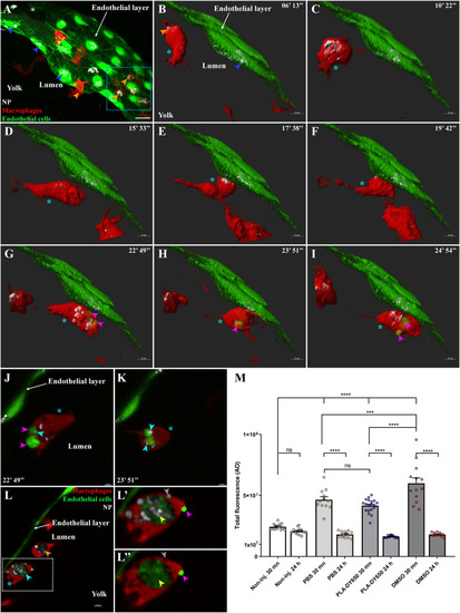
Transfer of PLA-NP from endothelial cells to macrophages. Representative live-acquisitions of 3 dpf mpeg1:mCherry and fli:GFP double transgenic zebrafish, 30 min after the intravenous injection of far-red fluorescent PLA-NP and using a spinning-disk confocal microscope. The acquisitions were made at the yolk circulation valley as illustrated in (A – 53 μm MIP, x63 objective), endothelial cells and macrophages being represented as a 3D surface reconstruction (green and red, respectively) and where PLA-NP (white) internalized by endothelial cells (blue arrowheads) and macrophage (orange arrowheads) can be observed. (B-I) The video (14 μm MIP), made across PLA-NP+ endothelial cells (blue arrowhead) of the endothelial layer covering the yolk, focuses on a macrophage (cyan star) which is already PLA-NP+ (orange arrowheads). At first, the macrophage enters in the proximity of the PLA-NP-positive endothelial cells (B-D). After almost 10 min at this position (E-F), GFP-labelled fragments of endothelial cell can be observed within the macrophage (G-I - magenta arrowheads) while the endothelial cells remain alive (J-K). Optical sections (1 μm) from G and H respectively, indicate the presence of PLA-NP (cyan arrowheads) inside the GFP-labelled fragment of endothelial cells (magenta arrowheads) that have been transferred to the macrophage (cyan star). (L) On an acquisition few minutes after the video, PLA-NP can be seen inside a GFP-negative macrophage (orange arrowhead), and within the GFP+ macrophage (cyan star and arrowhead). (L'-L") A close-up of the latter macrophage shows the presence of GFP-/PLA-NP+ cellular compartment (grey arrowhead), along with a massive GFP+/PLA-NP+ one in which both PLA-NP and the GFP+ cytosolic fragments of the endothelial cell co-localize (yellow arrowhead). Note the presence of a bright GFP+ structure at the surface of the macrophage (magenta arrowhead). (M) Analysis of the cell death following PLA-NP injection, assessed by the overall acridine orange fluorescence intensity (y-axis) of the whole zebrafish by stereomicroscopy. Intravenous injection of PLA-NP (DY650) does not induce cell death other than the mechanical injury from the injection, which is healed by 24 h post-injection. Each dot represents one single individual. Error bars: SEM. Significance level is indicated as: ***p < .001 and ****p < 0,0001. Number of fish per condition (30 min / 24 h): Non-injected (14 / 15), PBS-injected (12 / 12), PLA-DY650-injected (19 / 14), DMSO-injected (13 / 11). Scale bars: 20 μm (A), 5 μm (B-I,L) and 3 μm (J-K). (For interpretation of the references to colour in this figure legend, the reader is referred to the web version of this article.) |