
Fig. 1
- ID
- ZDB-FIG-210413-21
- Publication
- Rességuier et al., 2021 - Biodistribution of surfactant-free poly(lactic-acid) nanoparticles and uptake by endothelial cells and phagocytes in zebrafish: Evidence for endothelium to macrophage transfer
- Other Figures
- All Figure Page
- Back to All Figure Page
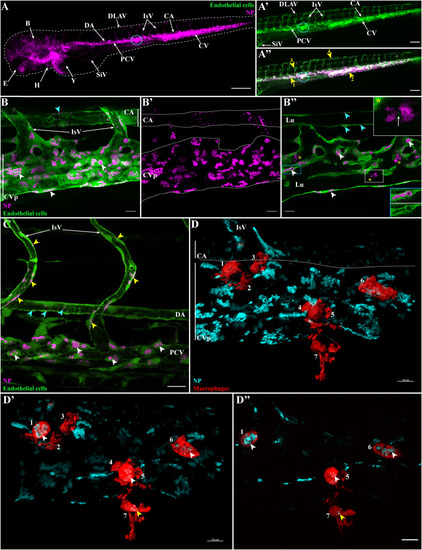
Internalization of PLA-NP by endothelial cells and macrophages. Representative live-acquisitions of 3 dpf zebrafish, 2 h after intravenous administration of PLA-NP, using a stereomicroscope (A-A") or a spinning-disk confocal microscope (B-D"). (A-A") Fluorescent PLA-NP (magenta) appear closely associated (yellow arrows) with the whole vasculature of fli:GFP+ fish (green). Injection sites are usually at the post-cardinal veins (blue circle) or within the inferior region of the caudal vein. As illustrated by the maximum intensity projection (MIP), which displays both PLA-NP and endothelial cells (B) and PLA-NP only (B′), strong accumulations of PLA-NP are observed within the endothelial cells of the caudal vein plexus (white arrowheads). PLA-NP are also taken-up by the endothelial cells of the caudal artery (cyan arrowheads). (B″) As revealed by optical sections (B), among both the caudal vein plexus and the caudal artery (white and cyan arrowheads, respectively), PLA-NP within endothelial cells are stored inside GFP negative cellular compartments. This localization is even more striking when the fluorescence signal from PLA-NP is removed (blue panel). Cluster of PLA-NP could be observed among luminal phagocytes (yellow stars). Co-internalization of GFP and PLA-NP within some luminal phagocytes has also been noticed (white arrow within white panel). (C) PLA-NP internalization is not restricted to the caudal vein plexus or the dorsal aspect of the post-cardinal vein. Endothelial cells from the whole post-cardinal vein (white arrowhead), the intersegmental vessels (yellow arrowhead) and the dorsal aorta (cyan arrowhead) are PLA-NP-positive, illustrating the pan-vascular internalization of PLA-NP. (D) From a mpeg1:mCherry zebrafish, MIP illustrating the presence of 7 macrophages (3D surface - red) close to PLA-NP (cyan) within the caudal vein plexus area. Both opening of the stack with a clipping plane (D') and the optic sections (D") reveal internalizations of PLA-NP by macrophage from the lumen (white arrowheads – 1, 5, 6) and outside the lumen (yellow arrowhead - 7). (B-B′) 17 μm MIP (x60 objective) from which (B″) is a 1 μm thick optical section. (C) 50 μm MIP (x60 objective). (D) 13 μm MIP (x60 objective) with a 3D surface reconstruction of the macrophage fluorescence signal. (D') illustrates an opening the MIP with a clipping plane, while (D") is a 1 μm thick optic section from the stack. Annotations: B, brain; CA, caudal artery; CVp, caudal vein plexus; DA, dorsal aorta; DLAV, dorsal longitudinal anastomotic vessel; E, eye; H, heart; IsV, intersegmental vessel; Lu, lumen; PCV, post-cardinal vein; SiV, sub-intestinal veins; Y, yolk. Scale bars: 200 μm (A), 100 μm (A'-A"), 20 μm (C) and 10 μm (B-B″, D-D"). (For interpretation of the references to colour in this figure legend, the reader is referred to the web version of this article.) |