
Fig. 3
- ID
- ZDB-FIG-210413-23
- Publication
- Rességuier et al., 2021 - Biodistribution of surfactant-free poly(lactic-acid) nanoparticles and uptake by endothelial cells and phagocytes in zebrafish: Evidence for endothelium to macrophage transfer
- Other Figures
- All Figure Page
- Back to All Figure Page
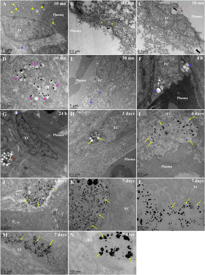
Fate of PLA-NP(Gold) once internalized by endothelial cells, from 10 min to 7 days post-injection, as observed with TEM. Representatives electron micrograph of ultrathin (60 nm) epon sections from zebrafish larvae injected at 3 dpf with PLA-NP encapsulating gold particles (<10 nm), 10 min (A-D), 30 min (E), 4 h (F), 24 h (G), 3 days (H), 4 days (I-J) and 7 days (K-N) post-injection. (A-D) 10 min following intravenous injection, PLA-NP are circulating within the bloodstream (A - yellow arrowhead) and internalized by endothelial cells (A - blue arrowhead). Internalization of PLA-NP by endothelial cells involve an invagination of the plasma membrane, resembling clathrin-mediated internalization (B - yellow stars), as well as figures resembling phagocytosis (C – red stars). Numerous NP were already being condensed within cellular compartment of some endothelial cells (D – magenta arrowheads). 30 min after the injection, no more PLA-NP could be observed within the bloodstream, in contrast to the massive concentration of PLA-NP within endothelial cells (E - blue arrowhead). No sign of degradation could be observed from PLA-NP internalized by endothelial cells (A, E-G - blue arrowhead) and underlying phagocytes (G - red arrowhead) during the first 24 h. Starting from 3 days post-injection, change in the colloidal stability of internalized PLA-NP was evident in some cells, encapsulated gold-particles being released from the NP-PLA matrix (H - yellow arrows). PLA-NP degradation was seemingly more pronounced 4 days post-injection, within some endothelial cells PLA-NP could are observed with a shrunken size (I - yellow arrows) while in others there are more gold crystals free rather than inside the PLA matrix (J - yellow arrows). Finally, a week after the injection different shade of PLA-NP degradation could be observed inside endothelial cells, cellular compartment filled with a paste of amorphous PLA containing gold crystals (K-L - yellow arrows), shrunken PLA-NP with released gold crystals (M - yellow arrows) and cellular compartment where only aggregated gold crystals remain (N - yellow arrows). Annotations: EC, endothelial cell. Scale bars: 1 μm (A,E,G), 500 nm (F,H,J-K,M), 200 nm (B-D,I,L) and 100 nm (N). (For interpretation of the references to colour in this figure legend, the reader is referred to the web version of this article.) |