
FIGURE
Fig. 3
- ID
- ZDB-FIG-210329-38
- Publication
- Iwasaki et al., 2020 - Development of the anterior lateral line system through local tissue-tissue interactions in the zebrafish head
- Other Figures
- All Figure Page
- Back to All Figure Page
Fig. 3
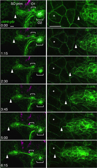
Migration of the SO pim. Images were selected from the time‐lapse recording of cldnb:gfp;atoh1a:rfp embryo around 30‐36 hpf (see Video S1). Right panels show higher magnifications of the leading edge of the primordium (see Video S2). Tip of the primordium with the landmark peridermal cell are indicated by arrowheads and asterisks, respectively. Lateral views, anterior is to the left. Scale bars: 20 μm |
Expression Data
Expression Detail
Antibody Labeling
Phenotype Data
Phenotype Detail
Acknowledgments
This image is the copyrighted work of the attributed author or publisher, and
ZFIN has permission only to display this image to its users.
Additional permissions should be obtained from the applicable author or publisher of the image.
Full text @ Dev. Dyn.