
Image
|
Figure Caption
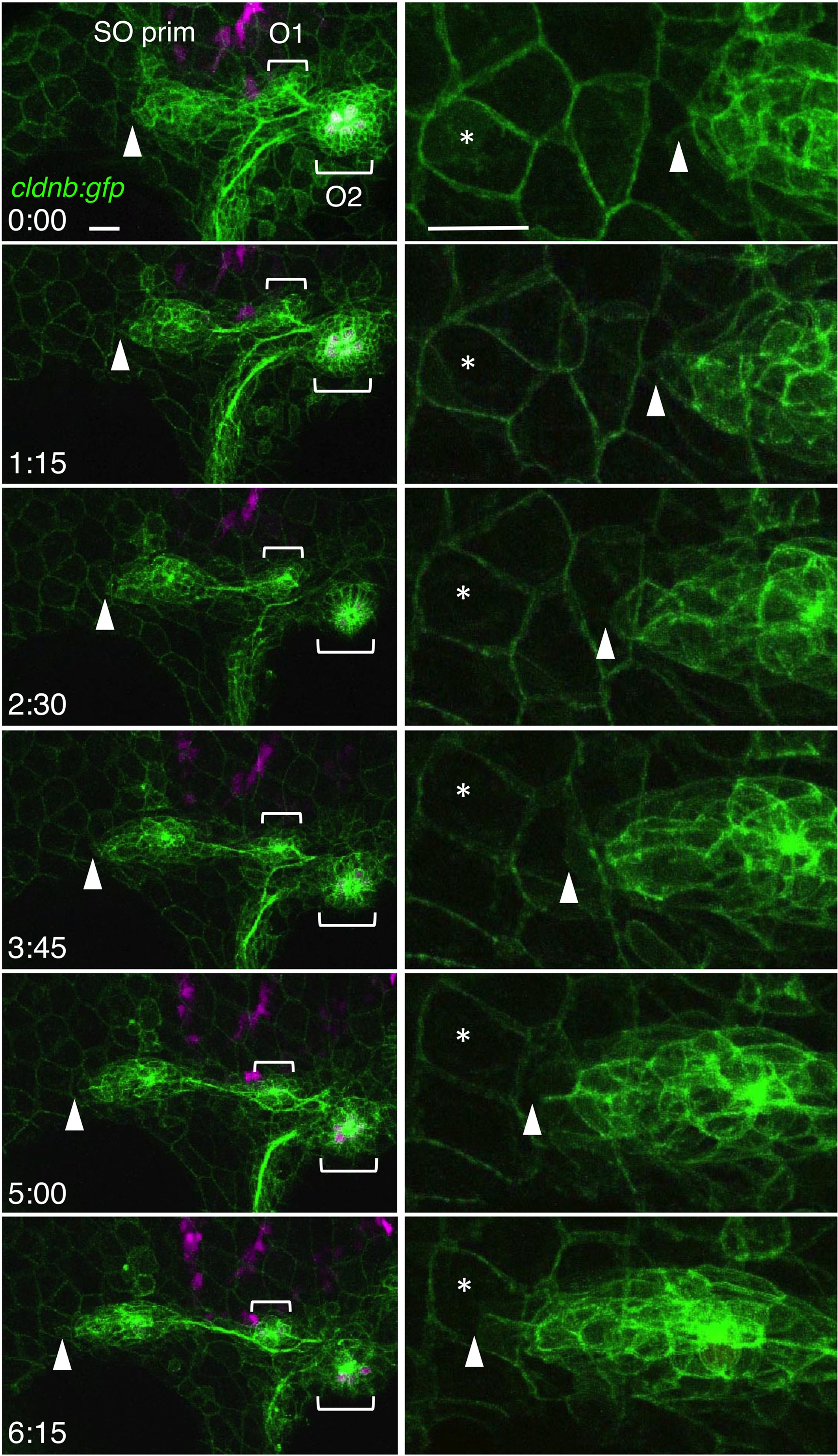
Fig. 3 Migration of the SO pim. Images were selected from the time‐lapse recording of cldnb:gfp;atoh1a:rfp embryo around 30‐36 hpf (see Video S1). Right panels show higher magnifications of the leading edge of the primordium (see Video S2). Tip of the primordium with the landmark peridermal cell are indicated by arrowheads and asterisks, respectively. Lateral views, anterior is to the left. Scale bars: 20 μm
Acknowledgments
This image is the copyrighted work of the attributed author or publisher, and
ZFIN has permission only to display this image to its users.
Additional permissions should be obtained from the applicable author or publisher of the image.
Full text @ Dev. Dyn.