
Fig. 2
- ID
- ZDB-FIG-210329-32
- Publication
- Heubel et al., 2020 - Endochondral growth zone pattern and activity in the zebrafish pharyngeal skeleton
- Other Figures
- All Figure Page
- Back to All Figure Page
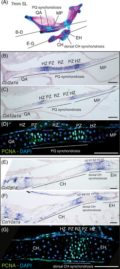
Molecular analysis of bidirectional endochondral growth zones in zebrafish. A, Section planes shown on alizarin red and alcian blue stained skeleton of 7 mm (SL) zebrafish mandibular and hyoid arches. B, Col2a1a expression in PQ synchondrosis and flanking QA and MP bones. C, Col10a1a expression in putative hypertrophic zones on either side of the PQ synchondrosis. D, anti‐PCNA immunostaining of presumptive proliferative and resting zone cells in PQ synchondrosis. E, Col2a1a expression in dorsal CH synchondrosis and flanking CH and EH bones. F, Col10a1a expression in putative hypertrophic zones on either sides of the dorsal CH synchondrosis. G, anti‐PCNA immunostaining of presumptive proliferative and resting zone cells in CH synchondrosis. Scale bar: 75 μm. CH, ceratohyal; EH, epihyal; HZ, hypertrophic zone; MP, metapterygoid; PZ, proliferative zone; PQ, palatoquadrate; QA, quadrate; RZ, resting zone |