
Fig. 1
- ID
- ZDB-FIG-210329-31
- Publication
- Heubel et al., 2020 - Endochondral growth zone pattern and activity in the zebrafish pharyngeal skeleton
- Other Figures
- All Figure Page
- Back to All Figure Page
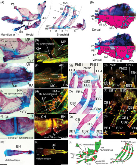
Endochondral growth zone locations in the zebrafish pharyngeal skeleton (13.5 mm SL). Bone stained with alizarin red, cartilage stained with alcian blue in panels A, B, C, E, G, I, K, M, O, Q. Old bone stained with calcein green, new bone stained with alizarin red in panels D, F, H, J, L, N, P, R. A, Anatomy of pharyngeal skeleton, including mandibular, hyoid, and branchial regions. B, Anatomy of neurocranial skeleton in dorsal and ventral views, with camera lucida outlines of bones in lower half of each image to facilitate identification. Synchondroses separating bones are colored in blue. C‐F, Mandibular skeleton regions. C, QA and MP bones across PQ synchondrosis. D, QA and MP growth. E, AR and RA bones and MC. F, AR growth. G‐L, Hyoid skeleton regions. G, HM and SY bones across HS synchondrosis. H, HM and SY growth. I, CH and EH bones across dorsal CH synchondrosis. J, CH and EH growth. K, BH bone and distal cartilage. L, BH growth. M‐R, Branchial skeleton regions. M, PhB1‐3 bones with cartilage pads and EB1‐4 bones with dorsal distal cartilages. N, PhB 1and 3 ossification, PhB2 growth and EB dorsal growth. O, EB1‐2 and CB1‐2 bones with distal cartilages. P, EB ventral growth and CB dorsal growth. Q, CB1‐2 bones with ventral distal cartilages and BB1‐3 bones across BB synchondroses. R, CB ventral growth and BB growth. S, Summary of locations of new bone (red) deposition detected in the pharyngeal arch cartilage‐derived skeleton. Scale bar: 140 μm. Dorsal mandibular arch: MP, metapterygoid, PA, palatine, PQ, palatoquadrate, QA, quadrate. Ventral mandibular arch: AR, articular; MC, Meckel's cartilage; RA, retroarticular. Dorsal hyoid arch: HM, hyomandibula; HS, hyosymplectic; IH, interhyal; SY, symplectic. Ventral hyoid arch: BH, basihyal; CH, ceratohyal; DHH, dorsal hypohyal; EH, epihyal; VHH, ventral hypohyal. Branchial arches: BB, basibranchial; CB, ceratobranchial; EB, epibranchial; HB, hypobranchial; PhB, pharyngobranchial. Neurocranium: BOC, basioccipital, E, ethmoid; EO, epioccipital; EOC, exoccipital; FR, frontal; LE, lateral ethmoid; OS, orbitosphenoid; PE, preethmoid; PRO, prootic; PS, parasphenoid; PTO, pterotic; PTS, pterosphenoid; SE, supraethmoid; SO, supraorbital; SOC, supraoccipital; SPH, sphenotic; VO, vomer |