
Fig. 5
- ID
- ZDB-FIG-210323-38
- Publication
- Wang et al., 2021 - Py3-FITC: a new fluorescent probe for live cell imaging of collagen-rich tissues and ionocytes
- Other Figures
- All Figure Page
- Back to All Figure Page
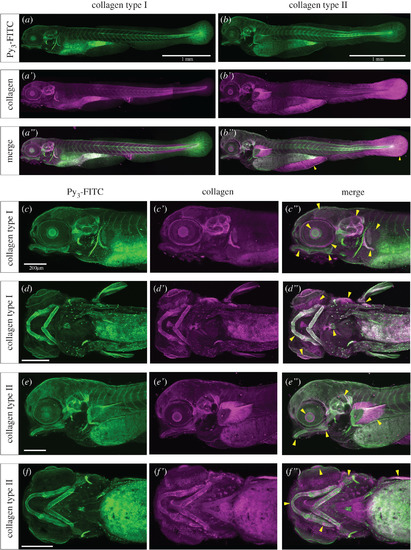
Py3-FITC can stain collagen type I- or II-positive tissues in zebrafish embryos. Reconstructed (a–b″,c–c″,e–e″) lateral views and (d–d″,f–f″) ventral views of the Py3-FITC and anti-collagen (a–a″,c–d″) type I and (b–b″,e–f″) type II antibody-stained zebrafish embryos at 5 dpf. (a″,b″,c″,d″,e″,f″) Py3-FITC-stained regions overlapped with collagen type I- or type II-positive regions. The yellow arrowheads represent Py3-FITC and collagen expression overlapping regions (b″,c″,d″,e″,f″). Enlarged views are shown of collagen (c′,d′) type I- and (e′,f′) type II-stained regions and their merged images (c″,d″,e″,f″). Scale bars in a,b, 1 mm; c,d,e,f, 200 μm. |