
Fig. 3
- ID
- ZDB-FIG-210323-36
- Publication
- Wang et al., 2021 - Py3-FITC: a new fluorescent probe for live cell imaging of collagen-rich tissues and ionocytes
- Other Figures
- All Figure Page
- Back to All Figure Page
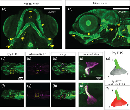
Cartilage tissues were stained with Py3-FITC. (a) Ventral and (b) lateral views of Py3-FITC-stained cartilage tissues 10 dpf. Meckel's (m), palatoquadrate (p), hyosymplectic (hs), ceratohyal (ch), scapulocoracoid (sco), cleithrum (cl), endoskeletal disc (ed) and opercle (op) were stained. (c–e) Ventral and (f–h) lateral views of Py3-FITC and Alizarin Red S double-stained zebrafish embryos 7 dpf. (i,j) Enlarged view of the yellow dotted squares shown in e, h. (k,l) Graphical illustration of Py3-FITC- and Alizarin Red S-stained opercles in i and j. Green indicates the Py3-FITC-stained region, and red indicates the Alizarin Red S-stained region of the opercles. The Py3-FITC-stained region was partially merged with the Alizarin Red-stained region. Parts of the opercle, joint apex (j), ventral (v), posterior (p) and joint socket (s) were visualized. Scale bars in a, b, c, 200 μm; i, 25 μm. |