
Fig. 4
- ID
- ZDB-FIG-210323-37
- Publication
- Wang et al., 2021 - Py3-FITC: a new fluorescent probe for live cell imaging of collagen-rich tissues and ionocytes
- Other Figures
- All Figure Page
- Back to All Figure Page
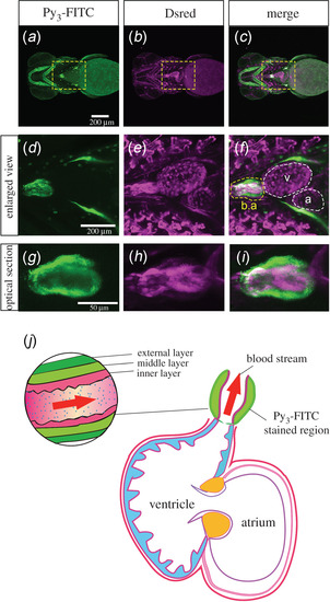
Bulbus arteriosus was stained with Py3-FITC. (a–c) Ventral view of the head and cardiomyocytes of Py3-FITC-stained Tg(fli:dsRed);casper zebrafish 7 dpf. (d–f) Enlarged views of the yellow boxed regions shown in a–c. Py3-FITC-stained b.a. but not the atrium (a) or ventricle (v) region. (g–i) Optical section views of Py3-FITC-stained and DsRed-positive regions in the b.a.. The graphical cross-sectional view of the zebrafish heart, which consists of the b.a., atrium and ventricle (j). The b.a. consists of inner and middle/external layers. Red arrows represent the direction of the bloodstream. The Py3-FITC-stained region is the middle/external layers of the b.a.. Scale bars in a, 200 µm; g, 50 µm. |